Research Article - Der Pharma Chemica ( 2018) Volume 10, Issue 6
Biochemical and Immunomorphometric Comparative Study Using Neem and Propolis Extracts For Hepatorenal Protection in Rats
Yassen NN1*, El-Sayed ME2, Hazaa MM3, El-Dougdoug KA4, Othman M3 and Elwan EE5
1Pathology Department, National Research Centre, Cairo, Egypt
2Plant Pathology Department, National Research Centre, Cairo, Egypt
3Microbiology Department, Faculty of Science, Benha, University, Egypt
4Microbiology Department, Faculty of Agriculture, Ain Shams University, Egypt
5Central laboratories, Ministry of Health, Egypt
Abstract
Background: Exposure to Alternariol or Patulin toxin is known to have toxic effects in different animal species, and to express toxicity in cells.
Purpose: The aim of this study is to compare the protective effect of Neem and Propolis extracts in hepatorenal toxicity caused by some mycotoxins.
Study design: Animals were divided into seven groups each of 5 animals and received single oral dose of 3 ml/rat, group 1, received A. alternata toxin; group 2, Alternaria alternate plus neem extract; group 3, A. alternata plus propolis extract; group 4, received Penicillium expansum toxin; group 5, P. expansum Patulin extract plus neem extract; group 6 P. expansum Patulin extract plus propolis extract; group 7, received normal water as a control.
Methods: ALT, Urea and Creatinine were detected in all groups after two weeks of feeding, the expression of Caspase-3 and iNOS in liver and kidney tissues were assessed immunomorphometrically by image analysis system.
Results: Animals received Alternariol or Patulin toxin alone showed significant changes in serum biochemical parameters and histological picture of the liver and kidney. Whereas, animals treated with Neem or Propolis extract in combination with Alternariol or Patulin toxins were comparable to control. The immunomorphometric parameter detected that the Propolis extract is more effective in the treatment especially with P. expansum toxin.
Conclusion: It could be concluded that the extract of Neem or Propolis induce its protective effect via increase the antioxidant capacity and the inhibition of lipid peroxidation.
Keywords
Alternaria alternate, Penicillium expansum, Neem extract, Propolis extract, Immunomorphometric parameters
Introduction
Apple (Malus domestica Borkh.) is one of the most important fruit species worldwide. Apple may get contaminated during growth, harvest, transportation and further processing and handling with microbes from soil, air, water or animal wastes [1]. Some moulds are related to a range of pathologies ranged from gastroenteritis to cancer, as these mycotoxins are highly toxic [2]. Some of them are responsible of the presence of mycotoxins in the crops (Alternaria, Claviceps and, Fusarium) and some of them in post-harvest processes (Aspergillus, Penicillium and Fusarium) [3].
The Alternaria genus produces more than 70 known mycotoxins and phytotoxins but only a few occur in food or are of major toxicological significance. Alternaria toxins which contaminate food are (not exhaustive): alternariol, alternariol monomethyl ether, tentoxin, tenuazonic acid, altertoxins, stemphyltoxin III, Alternaria alternate f. sp. lycopersici toxins [4]. Penicillium expansum, the blue mold that causes soft rot of apples, pears and other fruits, is recognized as one of the most common producers of the mycotoxin patulin. Patulin is quite toxic at high concentration in laboratory settings, but evidence for natural poisoning is indirect and inconclusive [5]. Several strategies are available for the detoxification of mycotoxigenic pathogens. These can be classified as physical, physicochemical, chemical, and biological approaches [6]. Biological control method is preferred because it is selective with no side effects, and is relatively cheap [7].
Neem (Azadirachta indica) is a tree in the mahogany family Meliaceae, is evergreen tree found in most tropical countries. Neem has anti-inflammatory, anti-tumor, immune-modulatory, anti-ulcer effects, hypoglycemic and other activities. These activities of Neem are due to presence of compounds like Nimbidin, Sodium nimbidate, Gedunin, Nembolide, Cyclic trisulphide, PolySaccharides, Polypeptidoglycan [8]. Due to its efficacy azadirachtin, a tetranortriterpenoid obtained from neem seeds, has emerged as a natural biopesticide [7].
Propolis is a substance collected by worker bees (Apis mellifera) from the bark of trees and leaves of plants. Propolis shows pharmacological activities, such as antifungal, antibacterial, anticancer or anti-inflammatory to name a few, among other activities, have attracted the researchers' interest [9].
Material and Methods
Mycotoxin production
Both A. alternata and Penicillium expansum were propagated as pure culture in 150 ml yeast extract sucrose (YES) containing 2% yeast extract and 15% sucrose / liter distilled water [10]. Each isolate was inoculated with 0.1 ml of a spore suspension containing approximately 106 spores’ ml-1 and incubated at 26 ± 2°C for 14 days.
Experimental animals
Three months old Sprague–Dawley female rats 100-120 g were used in the current study. They were purchased from the National Research Centre (NRC; Giza, Egypt). Animals received human care in compliance with the guidelines of the animal care and use committee of the NRC. Experiments were performed according to the National Regulations of Animal Welfare and Institutional Animal Ethical Committee (IAEC) and the national and international ethics guidelines stated by the ethics committee of NRC. Food consumption was approximately 15 g/female/day [11]. They were allowed to freely consume tap water and were fed according to the indicated experimental diets.
Experimental design, toxicity of mycotoxins and Biological control
Animals were divided into seven groups each of 5 animals, treated for two weeks as follows: group 1, received A. alternata toxin (Alternariol) as drink; group 2, treated orally with Alternaria alternate (Alternariol) extract plus neem extract; group 3, treated orally with Alternaria alternate (Alternariol) extract plus propolis extract; group 4, received P. expansum toxin (Patulin) extract; group 5, treated orally with P. expansum (Patulin) extract plus neem extract ;group 6, treated orally with P. expansum (Patulin) extract plus propolis extract; group 7, received normal water (untreated) as a control. The experimental determinations were made in rats fed on diets for two weeks with the same basic [12]. The animals were observed daily for signs of toxicity.
Blood samples
All animals were not fed on the day of the sacrifice. After the experimental period (14 days), the blood samples were collected from retro-orbital venous plexus for biochemical analysis in 15 ml-polypropylene tubes containing heparin as anticoagulant and centrifuged at 3000 rpm for 10 min to separate the plasma and used for biochemical serum analysis of kidney and liver function. Also, Blood draws (150-200 μl) were made using 1 ml syringes connected to the jugular cannula. Blood samples obtained from treated and untreated rats were analysis [13].
Biochemical evaluation of experimental animals
Determination of urea
Determination of urea level in serum using the enzymatic colorimetric method [14]. Using kit obtained from BioMérieax SA (France).
Determination of Creatinine
Determined in serum using commercial kits purchased from Stanbio Laboratory, Inc. (San Antonio, Texas, USA) [15].
Histopathological
Liver, Kidney specimens from all animals were dissected immediately after death, fixed in 10% neutral-buffered formal saline for 72 h. These specimens were washed in tap water for half an hour then dehydrated in ascending grades of alcohol, cleared in xylene and embedded in paraffin. Serial sections of 5 μm thick were cut, stained with Haematoxylin and eosin for histopathological investigation [16].
Immunohistochemistry for Caspase-3 and inducible Nitric Oxide Synthase
Paraffin-embedded sections were deparaffinized, and hydrated. Immunohistochemistry was performed with a mouse monoclonal caspase-3 and inducible nitric oxide synthase (iNOS) for detection of the caspase cleavage and iNOS activity. The paraffin sections were heated in a microwave oven (25 min at 720 W) for antigen retrieval and incubated with either anti-caspase or iNOS antibodies (1:50 dilution) overnight at 4°C. After washing with PBS, followed by incubation with biotinylated goat-anti-rabbit- immunoglobulin G secondary antibodies (1:200 dilution; Dako Corp.) and streptavidin/alkaline phosphatase complex (1:200 dilution; Dako) for 30 min at room temperature, the binding sites of antibody were visualized with DAB. After washing with PBS, the samples were counterstained with H&E for 2–3 min, and dehydrated by transferring them through increasing ethanol solutions (30%, 50%, 70%, 80%, 95%, and 100% ethanol). Following dehydration, the slices were soaked twice in xylen at room temperature for 5 min, mounted, examined, and evaluated by high-power light microscope [17].
Histological and immunohistochemical assessment studies
The studies are carried out on H&E, Caspase-3 and iNOS stained slides. Using high power as the objective lens with magnification of 20x and an eye lens with a magnification of 10x (total magnification 200x). Images were examined and photographed under a digital camera (Microscope Digital Camera DP70, Tokyo) and processed using Adobe Photoshop version 8.0.
Immunomorphometric analysis methodology
The morphometric analysis was performed at the Pathology Department, National Research Center using the Leica Qwin 500 Image Analyzer (LEICA Imaging Systems Ltd, Cambridge, England,) which consists of Leica DM-LB microscope with JVC color video camera attached to a computer system.
Detection of Caspase-3 and inducible Nitric Oxide synthase percentage area
The morphometric analysis is carried out on Caspase-3 and iNOS stained slides. The area percentage measured in 10 fields in each slide. We started by detection of the marker color, then the software form a binary image for the area of stained by the marker. This area was measured using an objective lens with magnification of 20x and an eye lens with a magnification of 5x (total magnification 100x) determined as an area per field in micrometer square, area fraction and area percentage by using the interactive measurement software of the system. The results appear automatically on the monitor in the form of a table with the total, mean, standard deviation, standard error, the minimum area and the maximum area measured.
Statistical analysis
Biochemical results
Data are subjected to statistical analysis of variance and means separation were done.
Statistical analysis for immunomorphometric results
Results are expressed as mean ± SE. Comparison of the values before and after the treatment were made by paired Student's t-test. P < 0.05 was considered to be statistically significant.
Results
Biochemical studied (serum analysis)
Changes in serum biochemical parameters caused by P. expansum and A. alternata toxins and their control were recorded in Table 1. The A. alternata toxin group was increased for all tested biochemical parameter compared with control group. No significant difference was found between biochemical parameters in animal received the two toxins alone. No significant difference was found between biochemical parameters in animal treated with two tested extracts plus A. alternata toxin while there was significance between the two tested extracts with P. expansum toxin. The present data Table 1 revealed that there were increased kidney function, Urea and Creatinine. Also AST and ALT which indicate the liver function increased in first and fourth group which received the toxins. The rest groups all their tested biochemical parameters were in normal limit.
| Groups | Urea mg/dl | Creatinine mg/dl | AST u/l | ALT u/l | LSD 5% between Groups | ||||
|---|---|---|---|---|---|---|---|---|---|
| D | R | D | R | D | R | D | R | ||
| Alternaria alternata | 43.08 a | 8.2 | 2.17 h | 97.3 | 20.7 d-f | 18.3 | 34.4 b | 7.5 | 25.099 A |
| A+NE | 34.74 ab | 12.7 | 0.92 h | 16.4 | 16.7 e-g | 4.6 | 20.9 d-f | 34.7 | 18.311 BC |
| A+Pro | 36.2 ab | 9.04 | 0.5 h | 54.5 | 12.2 g | 30.3 | 25.8 cd | 19.4 | 18.700 BC |
| Pencillium expansum | 42.9 a | 7.8 | 1. 7 h | 54.5 | 21.5 d-f | 22.9 | 34.1 bc | 6.6 | 25.064 A |
| P+NE | 35.5 ab | 10.8 | 0.8 h | 27.3 | 11.4 g | 34.9 | 19.2 d-g | 40 | 16.733 C |
| P+Pro | 36.8 ab | 7.5 | 0.7 h | 36.4 | 14.7 fg | 16 | 22.8 de | 28.8 | 18.794 BC |
| Control | 39.8 ab | - | 1.1 h | - | 17.5 d-g | - | 32 bc | - | 22.570 AB |
| LSD 5% between tests | 38.454 A | 1.124 D | 16.391 C | 27.043 B | - | ||||
D = Data analysis; R =Reduction%
Table 1: Effect of Penicillium expansum and Alternaria alternata fungi and their toxins on serum biochemical parameters in examined rat
Histopathological studies
Histopathological study of liver tissue
The histological examination revealed that liver tissue of the group which received A. alternata toxin alone Figure 1A-D or P. expansum toxin alone Figure 2A showing irritated hepatic tissue as the hepatocytes showing vesicular nuclei some with ballooning degeneration but still keeping their cytoplasmic wall, proliferating bile ducts epithelium (ductual hyperplasia) surrounded by inflammatory cells with activated kupffer cells. The group which received A. alternata toxin plus Neem extract and the other one which received A. alternata toxin plus Propolis extract both have been improved when compared with the control group Figure 1D. To be noticed that the group which treated with alternata toxin plus Neem extract has much better improvement Figure 1B, than the group which treated with A. alternata toxin plus Propolis extract which still have uncured hepatocytes in form of ballooning cytoplasm Figure 1C.
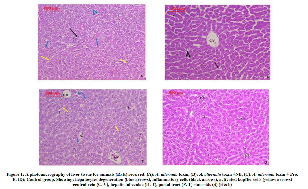
Figure 1: A photomicrography of liver tissue for animals (Rats) received: (A): A. alternata toxin, (B): A. alternata toxin +NE, (C): A. alternata toxin + Pro. E, (D): Control group. Showing: hepatocytes degeneration (blue arrows), inflammatory cells (black arrows), activated kupffer cells (yellow arrows) central vein (C. V), hepatic tabeculae (H. T), portal tract (P. T) sinusoids (S) (H&E)
There is an improvement in the group which received P. expansum toxin plus Neem extract Figure 2B as well formed hepatocytes which retained their cell wall some of them with deeply basophilic cytoplasm, while one which received P. expansum toxin plus Propolis extract Figure 2C has normal hepatic architecture when compared with the control group.
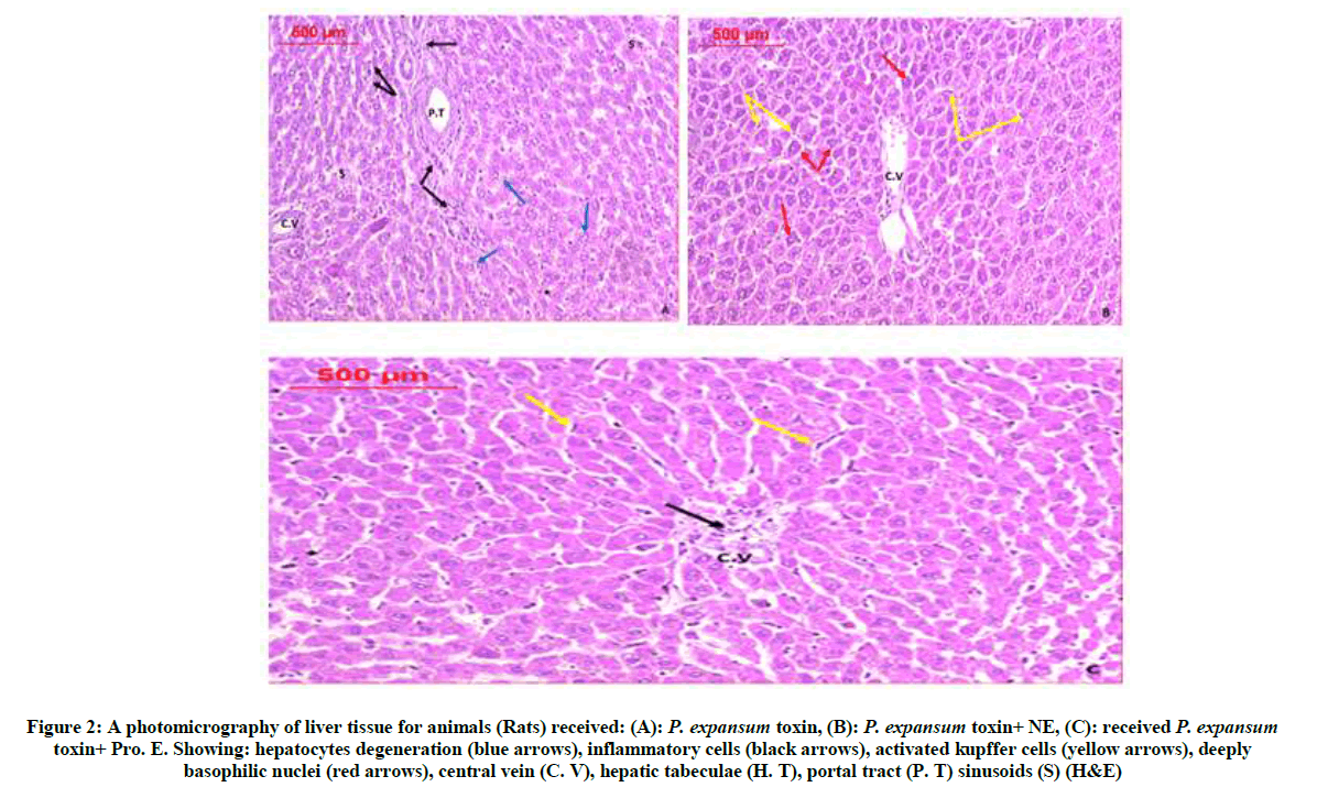
Figure 2: A photomicrography of liver tissue for animals (Rats) received: (A): P. expansum toxin, (B): P. expansum toxin+ NE, (C): received P. expansum toxin+ Pro. E. Showing: hepatocytes degeneration (blue arrows), inflammatory cells (black arrows), activated kupffer cells (yellow arrows), deeply basophilic nuclei (red arrows), central vein (C. V), hepatic tabeculae (H. T), portal tract (P. T) sinusoids (S) (H&E)
Histopathological study of kidney tissue
The renal tissue of the group which received A. alternata toxin alone Figure 3A-D or P. expansum toxin alone Figure 4A showing disturbed renal tissue as there are glomeruli destruction some are shrinked and degenerated, others have total lose of capillary tuft also most of the tubules have intratubular debris, losing their epithelial lining. These destructive changes have been improved in the groups which received A. alternata toxin plus Neem extract Figure 3B, and the one which received A. alternata toxin plus Propolis extract Figure 3C, as in the first one the renal tissue recovered as most of glomeruli retain its structure with normal Bowman`s capsule, minimal still with degenerated capillary tufts, the renal tubules have normal thickening, The second group has much better improvement as the renal tissue normally looking.
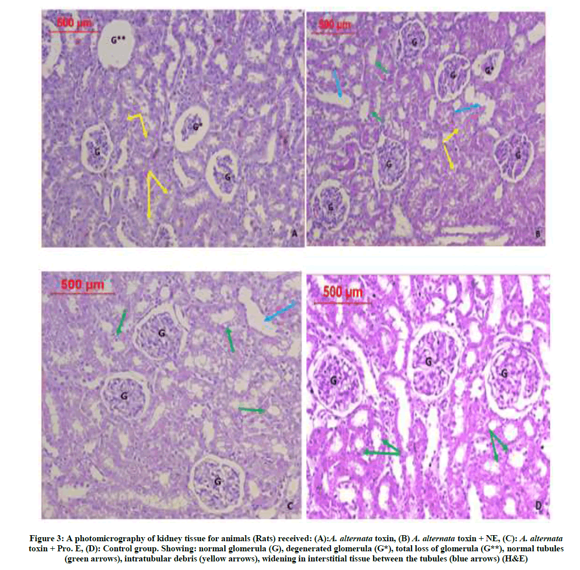
Figure 3: A photomicrography of kidney tissue for animals (Rats) received: (A):A. alternata toxin, (B) A. alternata toxin + NE, (C): A. alternata toxin + Pro. E, (D): Control group. Showing: normal glomerula (G), degenerated glomerula (G*), total loss of glomerula (G**), normal tubules (green arrows), intratubular debris (yellow arrows), widening in interstitial tissue between the tubules (blue arrows) (H&E)
The group which treated with P. expansum toxin plus Neem extract Figure 4B showing improved renal glomeruli but still others with shirked glomerular tufts, While the group which treated with P. expansum toxin plus Propolis extract Figure 4C showing more improvement as most of the glomeruli are looking normal, the tubules are minimally dilated but with intact healthy epithelium. All the previous groups are compared with the control group which received saline only Figure 3D.
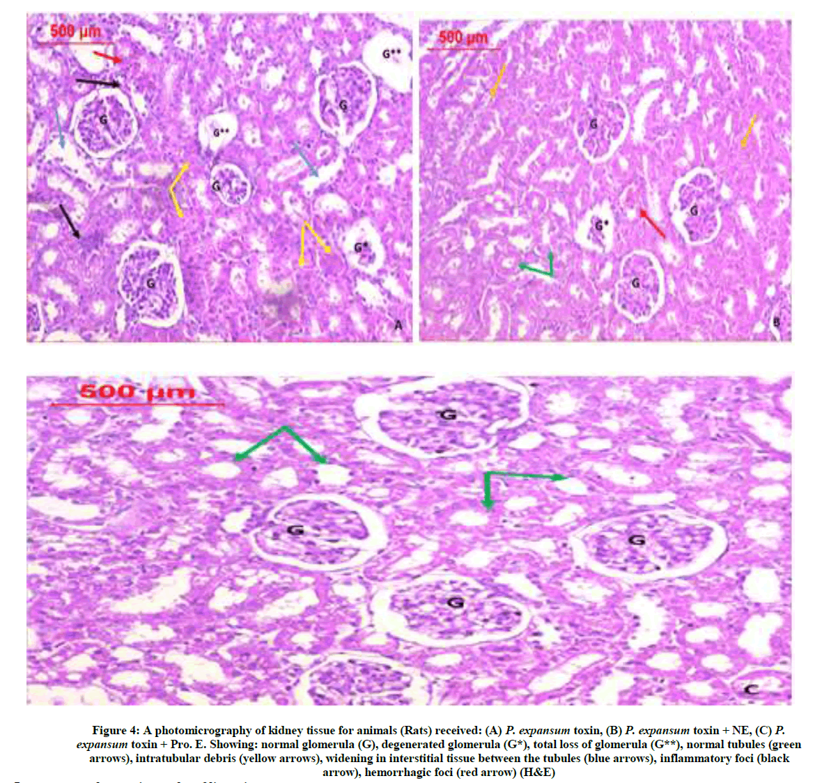
Figure 4: A photomicrography of kidney tissue for animals (Rats) received: (A) P. expansum toxin, (B) P. expansum toxin + NE, (C) P. expansum toxin + Pro. E. Showing: normal glomerula (G), degenerated glomerula (G*), total loss of glomerula (G**), normal tubules (green arrows), intratubular debris (yellow arrows), widening in interstitial tissue between the tubules (blue arrows), inflammatory foci (black arrow), hemorrhagic foci (red arrow) (H&E)
Immunomorphometric results of liver tissue
Detection and area percentage assessment of Caspase-3 in liver tissue
Figure 5A-G is a photomicrography of liver tissue showing IHC reaction of Caspase-3 & iNOS of all tested groups for further investigation morphometric measurement has been done. The percentage area of apoptotic cells in liver tissue for all studied groups Table 2 we noticed that the groups received the toxins A. alternata toxin and P. expansum toxin Figure 6A have the highest percentage areas of the positive cells. When these toxins treated with Neem extract in the groups of A. alternata toxin + NE Figure 6B and P. expansum toxin + NE have decreased the area percentage of immunoreactive positive cells denoting the improvement. On the other hand when we treated these toxins with Propolis extract as in groups of A. alternata toxin + Pro. E and P. expansum toxin + Pro. E Figure 6C having reducibility percentage of immunoreactive cells more than the groups treated with Neem extract which means that the Propolis extract is more effective in the treatment especially with P. expansum toxin Table 2.
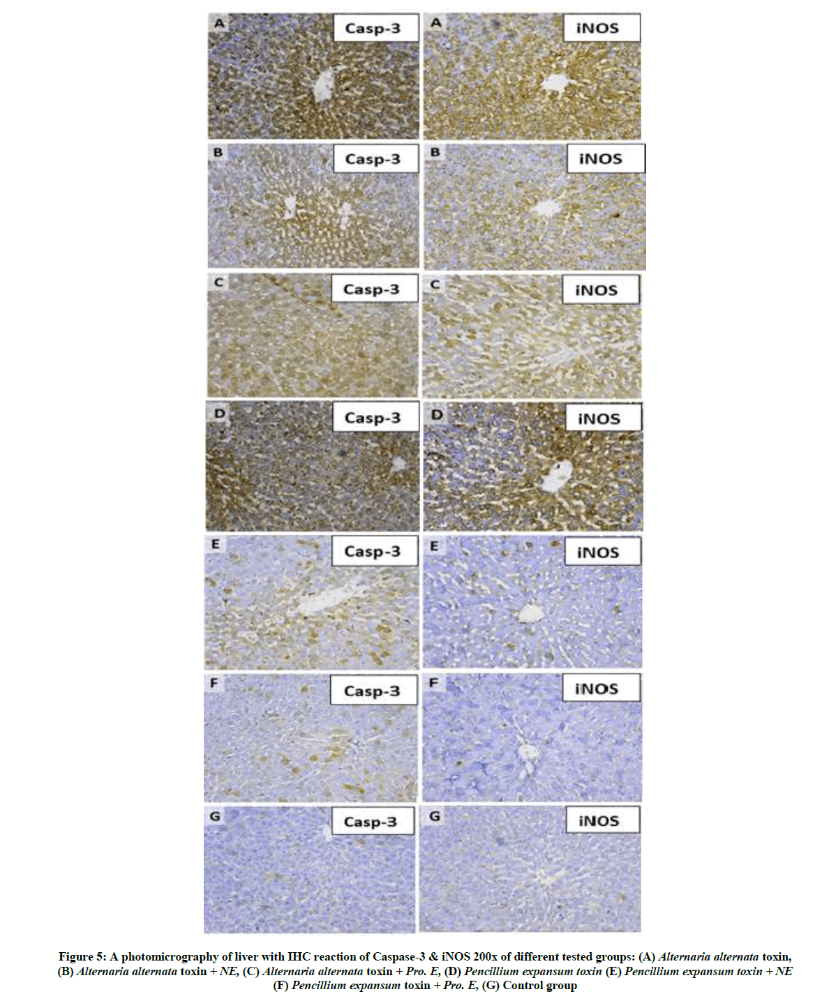
Figure 5: A photomicrography of liver with IHC reaction of Caspase-3 & iNOS 200x of different tested groups: (A) Alternaria alternata toxin, (B) Alternaria alternata toxin + NE, (C) Alternaria alternata toxin + Pro. E, (D) Pencillium expansum toxin (E) Pencillium expansum toxin + NE (F) Pencillium expansum toxin + Pro. E, (G) Control group
| Parameters | Groups | Area % of positivity in liver tissue | Area % of positivity in kidney tissue | ||
|---|---|---|---|---|---|
| Mean ± SE | Reduction % | Mean ± SE | Reduction %* | ||
| Alterneria alternata toxin | 22.16 ± 0.6 a | ------ | 27.81 ± 0.4 a | ------ | |
| A+NE | 8.45 ± 1.4 a,b | 61.86% | 10.67 ± 1.7 a,b | 61.63% | |
| A+Pro | 18.79 ± 0.8 a,b | 15.20% | 22.94 ± 1.3 a,b | 17.51% | |
| Pencillium expansum toxin | 31. 92 ± 1.3 a | ------ | 28.84 ± 0.5 a | ------ | |
| P+NE | 8.75 ± 1.6 a,c | 72.58% | 10.16 ± 1.6 a,c | 64.77% | |
| P+Pro | 7.01 ± 0.9 a,c | 78.03% | 6.91 ± 0.02 a,c | 76.04% | |
| Control | 6.89 ± 1.7 | ----- | 5.04 ± 1.4 | ------- | |
a Significantly different from normal negative control (Saline) at P<0.05;
b Significantly different from postive control A. alternata toxin at P<0.05;
c Significantly different from postive control P. expansum toxin at P<0.05;
* Reduction % from the toxin alone
Table 2: Results of immunomorphometric measurments of Caspase-3 to the tested groups in liver and kidney tissue. Data were expressed as mean ± SE. Statistical analysis was carried out by one-way ANOVA
Detection and area percentage assessment of iNOS in liver tissue
The inducible Nitric Oxide Synthase is an indicator of the oxidative stress due to reactive oxygen radicals. All Sections prepared from the liver tissue to show immune-reaction of iNOS in form of fine brown granules within the cytoplasm and cell wall of hepatocytes the groups Figure 5. We used it in calculating the percentage area of oxidative stressed cells in liver tissue for all tested groups shown in Figure 6. The increased iNOS activity was in the groups received the toxins A. alternata toxin Figure 6D and P. expansum toxin. When these toxins treated with Neem extract in the groups of A. alternata toxin + NE and P. expansum toxin + NE Table 3 have improved oxidative stress as decreased areas of iNOS activity. On the other hand when we treated these toxins with Propolis extract as in groups of A. alternata toxin+ Pro. E Figure 6E and P. expansum toxin plus Propolis extract Figure 6F having the higher reducibility percentage of oxidative stressed cells when compared with groups which treated by Neem extract which makes the Propolis extract has the upper hand in the treatment Table 3.
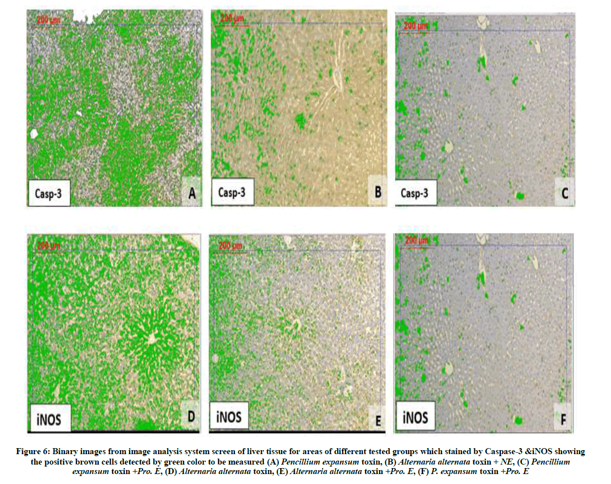
Figure 6: Binary images from image analysis system screen of liver tissue for areas of different tested groups which stained by Caspase-3 &iNOS showing the positive brown cells detected by green color to be measured (A) Pencillium expansum toxin, (B) Alternaria alternata toxin + NE, (C) Pencillium expansum toxin +Pro. E, (D) Alternaria alternata toxin, (E) Alternaria alternata toxin +Pro. E, (F) P. expansum toxin +Pro. E
| Parameters | Groups | Area % of positivity in liver tissue | Area % of positivity in kidney tissue | ||
|---|---|---|---|---|---|
| Mean ± SE | Reduction % | Mean ± SE | Reduction %* | ||
| Alternaria alternata toxin | 23.88 ± 1.9 a | ----- | 39.86 ± 1.7 a | ------ | |
| A+NE | 6.15 ± 1.2 a,b | 74.24% | 9.64 ± 1.7 a,b | 75.81% | |
| A+Pro | 5.52 ± 1.3 a,b | 76.88% | 3.72 ± 0.78 a,b | 90.66% | |
| Pencillium expansum toxin | 19.82 ± 0.9 a | ----- | 35.11 ± 1.1 a | ----- | |
| P+NE | 4.02 ± 1.5 a,c | 79.71% | 3.25 ± 1.02 a,c | 90.74% | |
| P+Pro | 3.24 ± 0.99 a,c | 83.65% | 2.81 ± 0.44 a,c | 91.99% | |
| Control | 1.62 ± 1.3 | ----- | 2.56 ± 0.63 | ------ | |
a Significantly different from normal negative control (Saline) at P<0.05;
b Significantly different from postive control A. alternata toxin at P<0.05;
c Significantly different from postive control P. expansum toxin at P<0.05;
* Reduction % from the toxin alone
Table 3: Results of immunomorphometric measurments of iNOS to the tested groups in liver and kidney tissue. Data were expressed as mean ± SE. Statistical analysis was carried out by one-way ANOVA
Immunomorphometric results of kidney tissue
Detection and Area percentage assessment of Caspase-3 in kidney tissue
Figure 7 showing IHC reaction of Caspase-3 & iNOS in kidney tissue for all tested groups, for further investigation morphometric measurement has been done. The morphometric measurement for the percentage area of apoptotic cells in kidney tissue for all studied groups Table 2, we found that the groups received the toxins A. alternata toxin and P. expansum toxin Figure 8A have the highest percentage areas caspase positivity. When these toxins treated with Neem extract in the groups of A. alternata toxin +NE Figure 7B and P. expansum toxin + NE have decreased areas of apoptotic cells. While when we treated these toxins with Propolis extract as in groups of A. alternata toxin +Pro. E and P. expansum toxin + Pro. E Figure 8C. Which means that the Propolis extract is more effective in the treatment especially with P. expansum toxin Table 2.
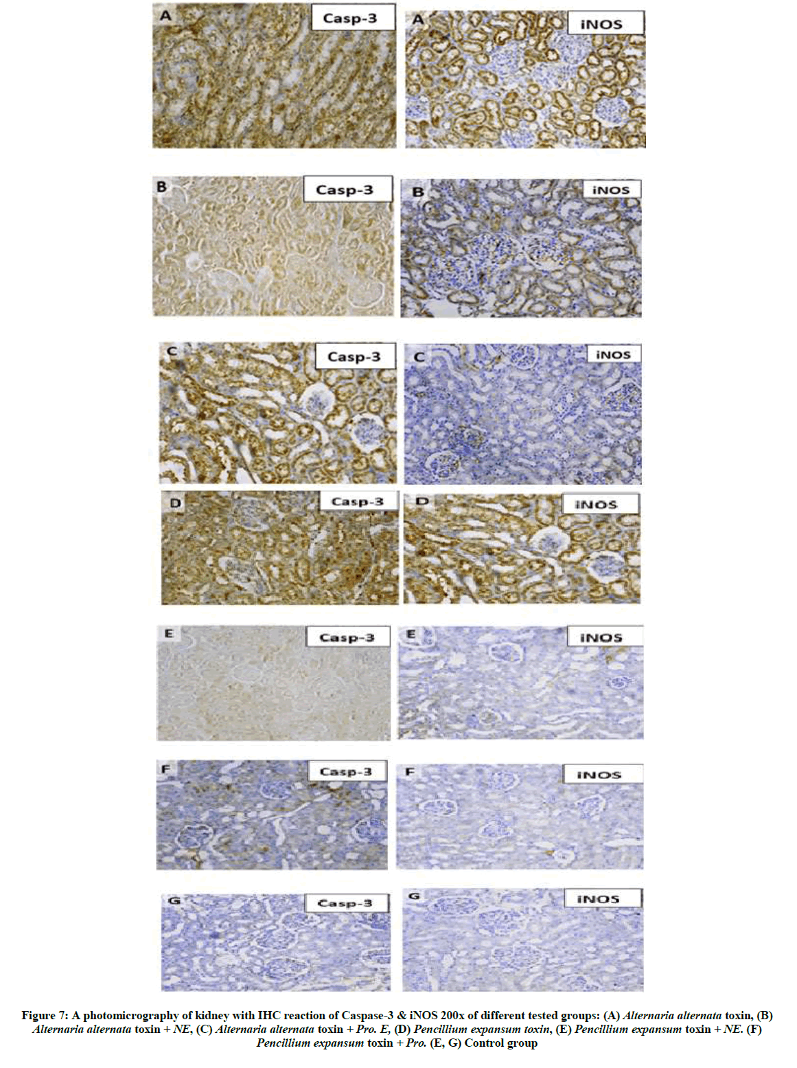
Figure 7: A photomicrography of kidney with IHC reaction of Caspase-3 & iNOS 200x of different tested groups: (A) Alternaria alternata toxin, (B) Alternaria alternata toxin + NE, (C) Alternaria alternata toxin + Pro. E, (D) Pencillium expansum toxin, (E) Pencillium expansum toxin + NE. (F) Pencillium expansum toxin + Pro. (E, G) Control group
Detection and Area percentage assessment of iNOS in kidney tissue
We used iNOS as an indicator of the oxidative stress in all tested groups due to reactive oxygen radicals also for the endothelial dysfunction as shown in Figure 7. The morphometric measurement of iNOS activity in kidney tissue for all tested groups showed that There were increased iNOS activity in the groups received the toxins A. alternata toxin Figure 8D and P. expansum toxin. When these toxins treated with Neem extract in the groups of A. alternata toxin +NE Figure 8E and P. expansum toxin + NE have decreased areas of iNOS activity. Using the Propolis extract as treatment in groups of A. alternata toxin + Pro. E and P. expansum toxin+ Pro. E Figure 8F which gives the Propolis extract the upper hand in the treatment. All previous groups have statistically significance difference with the control group Figure 8G.
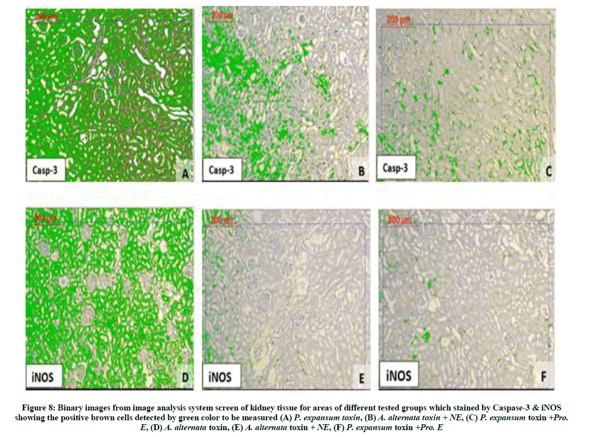
Figure 8: Binary images from image analysis system screen of kidney tissue for areas of different tested groups which stained by Caspase-3 & iNOS showing the positive brown cells detected by green color to be measured (A) P. expansum toxin, (B) A. alternata toxin + NE, (C) P. expansum toxin +Pro. E, (D) A. alternata toxin, (E) A. alternata toxin + NE, (F) P. expansum toxin +Pro. E
Discussion
Exposure to Alternaria or Penicillium toxin are known to have toxic effects, and to express toxicity in cells via the induction of oxidative stress. The obtained data indicated that A. alternata or P. expansum toxin alone increased significant all tested biochemical parameters but, showed non-significant increased with creatinin in serum of all treated groups compared with control group which was in normal limit.
El-Sawi et al. [18] found that, levels of AST and GGT were increased significantly in serum of all treated groups compared with control group but ALT was increased significantly in treated group after one week only. Propolis shows, in varying degrees, fungicidal effects against numerous species. The highest degree of inhibition observed, corresponded to a propolis concentration of 4% and the most affected microorganisms are A. alternata and Penicillium digitatum Abou El-Enain et al. [19] found that. Our results shown several changes in liver and kidney tissues represented by degeneration and necrosis in hepatocytes compared with control group. Whereas, the animals treated with Neem or Propolis extract alone or in combination with A. alternata or P. expansum toxins were comparable to the control. The same changes were obtained by Moss [20] and Al-Hazmi [21]. they reported that as the metabolism of Patulin mainly takes place in liver, leading to the events of histological changes represented by acute inflammation in hepatocytes, and swelling of the cells, causing relax and expand the blood vessels which lead to pooling of blood in central vein, vascular congestion, degeneration and necrosis in hepatocytes [22]. Awni et al. [23] found that, female rats received Apple juice contaminated with Patulin at 3 mg/rat had caused histological changes in the kidney and liver tissues represented by degeneration and necrosis in hepatocytes and had increased with increasing of repeated doses of toxin.
In fact, in recent studies, propolis has been demonstrated to play an important role in preventing the oxidative stress, apoptosis and necrosis. Possible mechanisms of hepatoprotective action of propolis extract may be due to its free radical scavenging activity as indicated by decrease in lipid peroxidation and increase in glutathione contents. Improved enzymatic biochemical parameters and histopathological observations also indicated recovered structural and functional integrity of the hepatorenal cells and provided further support to the proposed protective mechanism of action by propolis. Thus, it can be concluded that propolis extract possesses therapeutic potential against hepatorenal damage [24]. Our results were in harmony with those of Ayman et al. [25] who postulated that, propolis play an important role in preventing the retinal damage.
The protective effect of neem leaves extract may be due to ability for regulation the immune mediated through the suppressive effect on the production of important inflammatory mediators. Also, may be due to the capacity of catehins to scavenge free radicals and protect against oxidant stress induced by acrylamide. Neem fruits caused decreased serum activities of alkaline phosphatase and an improvement of ALT, AST and bilirubin values [26]. The non-hepatotoxic nature of neem was proved in the study performed by Haque et al. [27] who found unaltered and normal activities of serum ALT, AST, ALP as well as retained architecture of liver after neem treatment. Yanpallewar et al. [28] reported the hepatoprotective role of neem leaves extract against paracetamol-induced hepatic damage in albino rats.
Conclusion
It can be concluded that treatment with Neem or Propolis extract attenuated Alternaria and/or Penicillium toxicity in rats, preventing reduced LDH and the decrease in transaminases leakage to serum, which may play protective role against mycotoxin-mediated liver injury. Another mechanism for the protective role of the extract components is the enhancement of glutathione production which consider the first defense line in protecting the cell from oxidative stress.
References
- E. Chen, S.C. Ekker, Curr. Pharm. Biotechnol., 2004, 5(5), 409-413.
- M.S. Adams, M.C. Moss, Food microbiology, 2nd (Edn.), Royal Society of chemistry. Thomas Graham house, Science Park., Milton Road Cambridge CB 40 WF, UK, 2000.
- P. Ester, V.M. Liliana, G.C. Jesús, L. Joan-Maria, Rec. Adv Pharm Sci., 2013, 3, 131-144.
- J. Alexander, D. Benford, A. Boobis, C. Sandra, B. Cottrill, C. Jean-Pierre, A. Di Domenico, D. Doerge, E. Dogliotti, L. Edler, P. Farmer, M. Filipič, J. Fink-Gremmels, P. Fürst, T. Guérin, H.K. Katrine, M. Machala, A. Mutti, S. Josef, M. Rose, R. Leeuwen, EFSA Journal., 2011, 9(10), 2407.
- I.T. Dimitrios, D. Myrto, P.A. Polymnia, C.T. Eleftherios, Phytopathologia. Mediterranea., 2012, 51(1), 158-174.
- B.M. Gardener, D.R. Fravel, Biological control of plant pathogens: Research, Commercialization, and application in the USA, Plant Health Progress, 2002.
- A.A. Olufemi, O.A.T. Joseph, F A. Grace, G. J. B. A. H. S., 2014, 3(1), 106-109.
- B. Kaushik, I. Chattopadhyay, R.K. Banerjee, U. Bandyopadhyay, Current Sci., 2002, 82(11), 1336-1345.
- A. Mavri, H. Abramovic, T. Polak, J. Bertocelj, P. Jamnik, S.S. Mozina, B. Jersek, Chem. Biodivers., 2012, 9, 1545-1558.
- M.I. Ammar, M.A. El-Naggar, Int. J. Adv. Res., 2014, 2(4), 1216-1227.
- J. Liebert, I. Matławskab, W. Bylka, M. Murias, Environ. Toxicol. Pharmacol., 2006, 22, 58-63.
- S.E. Madrigal, B.E. Madrigal, M.R. Ma´rquez, A. Reyes, Food. Chem. Toxicol., 2006, 44, 2058-2063.
- N.G. Mangano, V.M. Cutuli, A. Caruso, E. De Bernardis, M. Amico-Roxas, Eur. Rev. Med. Pharmacol. Sci., 2001, 5, 1-6.
- D. Barham, P. Trinder, Analyst., 1972, 97(151), 142-145.
- H. Bartels, M. Bohmer, C. Heirli, Clin. Chem. Acta., 1972, 37, 193-197.
- N. Omar, The Egyptian Journal of Hospital Medicine., 2012, 48, 357-367.
- A.M. Gown, M.C. Willingham, J. Histochem. Cytochem., 2002, 50(4), 449-454.
- N.M. El-Sawi, H.M. Gashlan, S.H.H. Younes, R.F. Al-Massabi, S. Shaker, Life Science Journal, 2012, 9(4), 1143-1153.
- H.T.A. El-Enain, M.F.A. Rahman, K.A.M.A Elyousr, Ass. Univ. B ull. Environ. Res., 2009, 12(2), 99-106.
- M.O. Moss, J. Appl. Microbiol., 2008, 104, 1239-1243.
- M.A. Al-Hazmi, Toxicol., Health, 2012, 30(4).
- I. Alves, N.G. Oliveira, A. Laires, Mutagenesis., 2000, 15, 229-234.
- A.M. El-Sayed, M. Embaby, N.N. Yassen, M.M. Abdel-Galil, Heba, I. El-Gendy, Middle East J. Appl. Sci., 2015, 5(4), 1007-1016.
- E.M. Thanaa A, M.E. Ashraf, A.E. Nagla, J. Evol. Biol. Res., 2011, 3(1), 4-11.
- A.E.M.A.E. Kenawy, H.E.H. Osman, M.H. Daghestani, Environ. Toxicol., 2014.
- S.J. Boeke, M.G. Boersma, G.M. Alink, J.J. van Loon, A. van Huis, M. Dicke, I.M. Rietjens, J. Ethnopharmacol., 2004, 94, 25-41.
- E. Haque, I. Mandal, S. Pal, R. Baral, Immunopharmacol. Immunotoxicol., 2006, 28, 33-50.
- S.U. Yanpallewar, S. Sen, S. Tapas, M. Kumar, S.S. Raju, S.B. Acharya, Phytomedicine., 2003, 10, 391-396.


