Research Article - Der Pharma Chemica ( 2024) Volume 16, Issue 2
Curcumin and Ascorbic Acid Therapeutic Benefits in a Female Wistar Rat with Renal Tissue UVB-Induced Hyperthyroidism: A Histological and Biochemical Study
Gayatri Rai, Shashank Shakyawal, Narendra Namdev and Payal Mahobiya*Payal Mahobiya, Department of Zoology, Dr. Harisingh Gour University, Madhya Pradesh, India, Email: 1607payal@gmail.com
Received: 12-Mar-2024, Manuscript No. Dpc-24-129305; Editor assigned: 15-Mar-2024, Pre QC No. Dpc-24-129305 (PQ); Reviewed: 29-Mar-2024, QC No. Dpc-24-129305; Revised: 01-Apr-2024, Manuscript No. Dpc-24-129305 (R); Published: 29-Apr-2024, DOI: 10.4172/0975-413X.16.2. 249-256
Abstract
Renal issues are a major problem for humanity. The renal system may sustain damage from the potentially dangerous radiation. Therefore, the purpose of this study is to evaluate how well curcumin and ascorbic acid protect kidney tissue from being altered by UVB-induced hyperthyroidism in female Wistar rats. Twenty-four female’s sexually older Wistar rats weighing 130 g-150 g and aged 12-16 weeks were arbitrarily divided into four groups. The control group received normal food and water ad libitum. The UVB group was exposed to a dose of 280 nm of UVB radiation for 2 h daily. The UVB+ curcumin group received a dose of 280 nm of UVB radiation for 2 h daily and also an oral dose of curcumin (25 mg/kg body weight) daily. The UVB+ ascorbic acid group received a dose of 280 nm of UVB radiation for 2 h daily and also an oral dose of ascorbic acid (250 mg/kg body weight) daily. All the treatments last for 15 consecutive days. Body and kidney weight were measured. The histology of kidney tissue was done by H and E, PAS, MBB and Sudan black B staining and observed. The biochemical changes were measured by particular methods. UVB radiation caused histological and biochemical changes in Wistar rats, while curcumin and ascorbic acid protected these changes, with ascorbic acid showing greater protection. Curcumin and ascorbic acid at low doses can alleviate changes made in histology and biochemical reactions in cells induced by UVB radiations in female Wistar rats.
Keywords
Radiations; Curcumin; Ascorbic acid; Enzymatic and non-enzymatic assay; Histology
Introduction
Toxicants are substances that can be poisonous and may be man-made or naturally occurring. Different types of toxicants can be found in air, soil, water or even in food. In today’s changing global scenario, radiation is considered the most potent cause of oxidative stress mediated free radical flux which induces severe damage at various hierarchical levels in the living organisms. This flux interferes with oxidation/reduction based physiological mechanisms of all major cellular components (including water, lipid, proteins, sugars and nucleic acids) existing inside every organ of the body system [1]. The radiation effects can be influenced by various factors such as age, gender, exposure time and affected tissue. Thus, until today it has not been possible to precisely determine a threshold and linearity of low radiation dose effects, which makes it difficult to establish a safety limit for human health [2]. Radiations come in the category of systemic toxicants that can affect the entire body at a single time. These radiations have a vital effect on the body. The ionizing radiations and non-ionizing radiations are the types of radiations. Exposure to these radiations causes cellular damage and alteration in physiological and biochemical reactions in living cells. These radiations emit and transmitted by different sources which are absorbed by the animal body. UV radiations are non-ionizing radiations that have wavelengths ranging from 200 nm to 400 nm. These UV radiations are present in sunlight and have that much energy by which it can easily penetrate body cells and causes alteration in chemical and biological activities. As the cells or tissues are exposed to UV radiation, the water molecules undergo dissociation and produce free radicals and related species in the form of ROS. These, in turn, can act on biomolecules such as DNA, lipids and proteins and cause oxidative damage [3-4]. This non-ionizing radiation is categorized into three categories i.e., UVC, UVB and UVA having wavelengths of 200 nm-280 nm, 280 nm-320 nm and 320 nm-400 nm respectively [5-6]. The kidney is among the more radiosensitive late responding critical organs. Irradiation of both kidneys to a modest dose has resulted in nephropathy with arterial hypertension and anemia and the radiation damage develops slowly and may not become evident for months. Parts of one or even both kidneys can receive much higher doses [7]. Exposure to electromagnetic radiation in rat kidneys increased total kidney volume and decreased the number of glomeruli and induced renal toxicity [8-9]. Long-term exposure to electromagnetic radiation generates oxidative stress in rat kidneys and vitamin-c cures oxidative damage in renal tissue [10-11]. Although having much lesser penetrating power, UVB radiations are potent enough to cause DNA damage [12].
The present study is new in its field, which for the first time demonstrates the effect of repetitive UVB radiation induced hyperthyroidism on the kidney of female Wistar rats and we also investigated the protective effect of antioxidants (ascorbic acid and curcumin) against the morphological changes in cells induced by UVB radiation.
Materials and Methods
Chemicals
The chemicals and reagents used were of analytical grade. Ascorbic acid was purchased from Sigma-Aldrich Co., USA. Curcumin, NBT, NADPH, methionine and reduced glutathione were obtained from himedia, India and thiobarbituric acid, haematoxylin, eosin and the rest of the chemicals used were obtained from central drug house (P) Ltd, New Delhi, India.
UVB irradiations
UVB light (TL 20W/01 UVB narrowband made in Germany), which emitted UVB in the range of 280 nm (UVB), was used as the source to irradiate the Wistar rats. The irradiance was two hours daily for 15 days [13].
Experimental animals and treatment
Female adult Wistar rats weighing 130 g-150 g and aged 12-16 weeks were purchased from the college of veterinary sciences and animal husbandry Mhow (22.55º N, 75.75º E, M.P), India. All animals (n=24) were housed in plastic cages and were fed on the standard laboratory of diet daily food and water ad libitum. Rats were kept under laboratory conditions, with standard temperature (20°C ± 2°C) relative humidity of (50% ± 5%) and 12 h/12 h light and dark cycle. The experimental rats were randomly divided into four groups, with six rats in each group (Table 1).
| Groups name | Dose | Diet | Duration |
|---|---|---|---|
| Control | No treatment | Normal food and water | 15 days |
| UVB-induced | 280 nm of UVB radiation for 2 hours | Normal food and water | 15 days |
| UVB-induced+curcumin | 280 nm of UVB radiation for 2 hours+curcumin (25 mg/kg body weight) | Normal food and water | 15 days |
| UVB-induced+ascorbic acid | 280 nm of UVB radiation for 2 hours+ascorbic acid (250 mg/kg body weight) | Normal food and water | 15 days |
Table 1: Experimental design.
Ethical statement
The study was permitted by the department of pharmaceutical sciences Dr. Harisingh Gour Vishwavidyalaya (A central university) Sagar (M.P.), India (registration No. 379/Go/ReBi/S/01/CPCSEA) and all the international guidelines were followed for the care and use of laboratory animals.
Sample collection
At the end of the experiment, the animal was deadened with chloroform and blood samples were collected by cardiac puncture. One part of the blood was taken for whole blood parameter measurement while the other part was taken for hormonal estimation and biochemical analysis. Kidneys were dissected out, washed in ice-cold phosphate buffer saline and stored at -20C for further analysis.
Measurement of body and kidney weight
Body and kidney weight measurements were done with an electronic balance (Sartorius, BP210 S). Bodyweight was measured before and after the experiment. Kidney weight was measured after the experiment.
Histochemical examinations
Whole-body perfusion was done through the heart using 0.02 M Phosphate-Buffered Saline (PBS) and 4% PFA [14]. The kidney was removed and kept in the fixative for 48 h. Fixed tissues were dehydrated and then processed and cut 5 μm sections from paraffin-embedded kidneys and stained with haematoxylin and eosin, Periodic Acid Schiff (PAS) [15], Mercuric Bromophenol Blue (MBB) [16], Sudan black B [17] Images were analyzed under the microscope on 100X and 400X magnifications.
Tissue homogenization
For the biochemical examinations, kidney tissues were firstly homogenized in 0.02 M Tris-Cl pH 7.4 and prepared the 10% homogenate. The homogenate was centrifuged at 12500 rpm for 30 min at 4ºC. The supernatant was collected and stored at -20ºC for further analysis and protein estimation was performed [18].
Biochemical examinations
Serum urea and creatinine: Serum urea and creatinine values were determined with the commercial kits by an auto analyzer.
Enzymatic and non-enzymatic activity assessment: The lasting part of testis was used for analysis of lipid peroxidation [19], catalase [20], superoxide dismutase [21], GSH [22] levels in all the above groups.
Statistical analysis
All statistical investigation was performed by using a one-way analysis of variance. The data were uttered as mean ± SE. Dunnett test was used for the assessment between the control group and each treated group individually. P<0.05 was considered statistically different.
Results
Body and organ weight
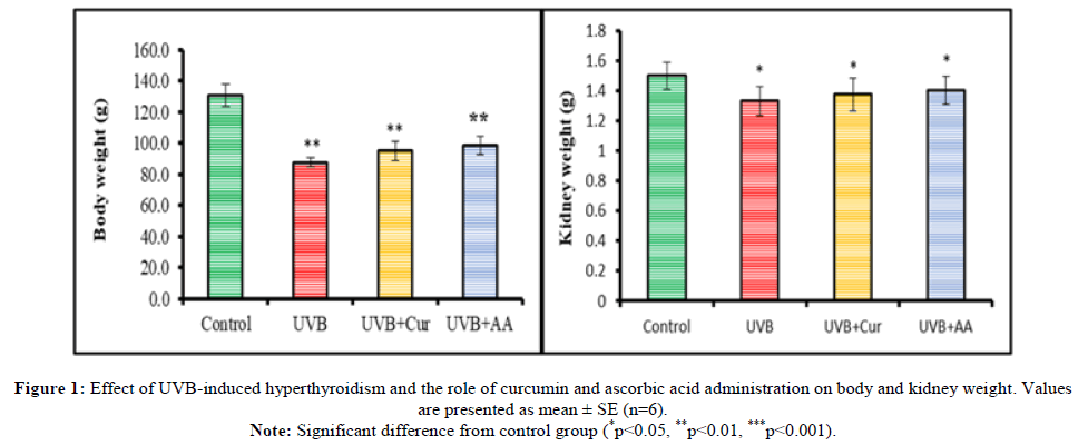
A significant reduction (p<0.01) in body and kidney weight of the UVB-induced hyperthyroidism group was shown as compared to the control group. The body and kidney weight of the Curcumin (Cur) and Ascorbic acid (AA) treated group was slightly significantly increased as compared to UVB induced hyperthyroidism group (Figure 1).
Note: Significant difference from control group (*p<0.05, **p<0.01, ***p<0.001).
Serum urea and creatinine estimation
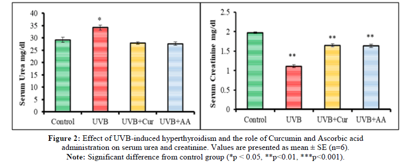
UVB-induced hyperthyroidism group showed a significant (p<0.05) elevation in serum urea as compared to the control group. Administration of curcumin and ascorbic acid showed a reduction of serum urea as compared to the UVB-induced hyperthyroidism group. It was shown no significant effect as compared to the control group. Serum creatinine showed a significant (**p<0.01) reduction in the UVB-induced hyperthyroidism group. Administration of curcumin and ascorbic acid showed a significant elevation of the UVB+Cur and UVB+AA groups (Figure 2).
Note: Significant difference from control group (*p < 0.05, **p<0.01, ***p<0.001).
LPO estimation
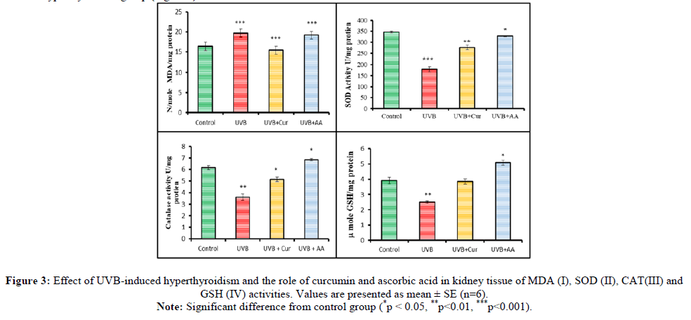
UVB-induced hyperthyroidism group showed a significant (***p<0.001) elevation of MDA levels in the kidney as compared to the control group. Administration of curcumin and ascorbic acid in UVB+Cur and UVB+AA treated groups (**p<0.01) showed a significant reduction of MDA level as compared to UVB induced hyperthyroidism group.
SOD estimation
The SOD activity was observed in the UVB-induced hyperthyroidism group was significantly reduced as compared to the control group. Administration of curcumin and ascorbic acid in UVB+Cur and UVB+AA treated groups; the activity of SOD was significantly (**p<0.01) elevated as compared to UVB induced hyperthyroidism group.
Catalase activity
UVB-induced hyperthyroidism group showed a significant (*p<0.05) decrease in catalase activity as compared to the control group. Administration of curcumin and ascorbic acid in UVB+Cur and UVB+AA treated group; the catalase level was shown to be increased as compared to UVBinduced hyperthyroidism group.
GSH activity
The GSH activity was significantly lower in the UVB-induced hyperthyroidism group as compared to the control group. Administration of curcumin and ascorbic acid in the UVB+Cur and UVB+AA treated group was significantly higher in GSH activity as compared to the UVBinduced hyperthyroidism group (Figure 3).
Note: Significant difference from control group (*p < 0.05, **p<0.01, ***p<0.001).
Histochemical analysis
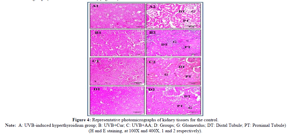
The normal histological appearance of glomerulus and tubular structure was observed in the control group (A), while in the UVB-induced hyperthyroidism group (B) degenerative, vascular changes and disappearance of the glomerular, bowman’s capsules and elongated proximal and convoluted tubular structure were seen. Epithelial cells of both proximal and distal convoluted tubules in the kidney had pyknotic nuclei and were damaged and desquamated. Co-administration of curcumin (UVB+Cur) and ascorbic acid (UVB+AA) showed less damage to kidneys, as compared to UVB-induced hyperthyroidism in rats. However, repaired degenerative epithelial cells and positive vascular changes were observed in coadministrative groups (UVB+Cur and UVB+AA) (Figure 4).
Note: A: UVB-induced hyperthyroidism group; B: UVB+Cur; C: UVB+AA; D: Groups; G: Glomerulus; DT: Distal Tubule; PT: Proximal Tubule) (H and E staining, at 100X and 400X, 1 and 2 respectively).
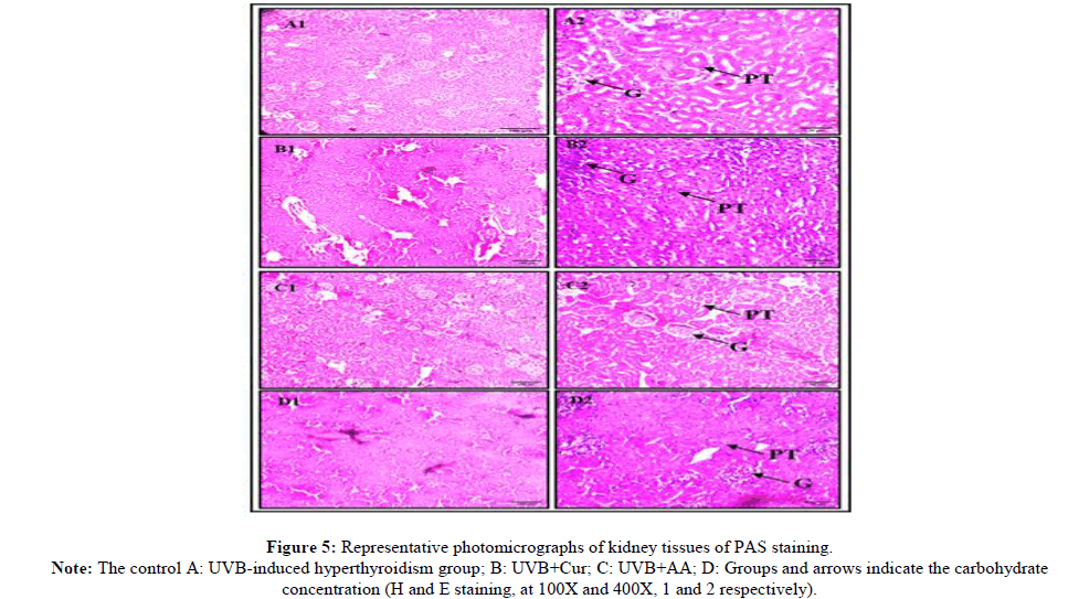
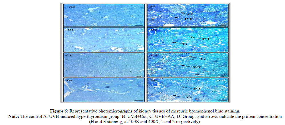
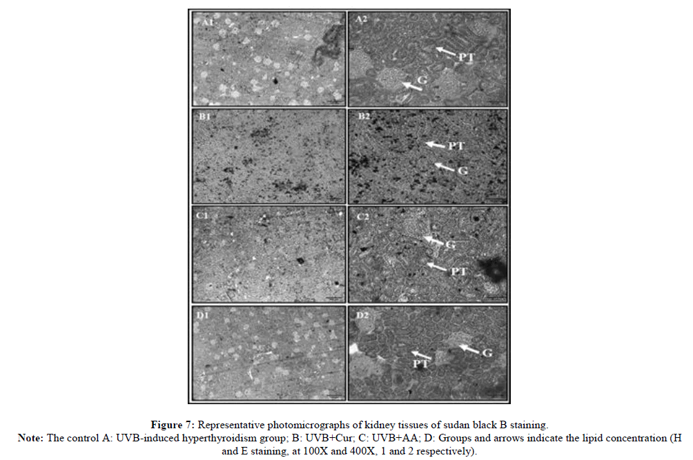
In histochemical analysis, the glomerular vacuoles were negative for performance acid-Schiff and positive for PAS (Figure 5), MBB (Figure 6) and Sudan black B (Figure 7).
Histological examination of kidneys of control group rat stained with periodic acid-Schiff showed the positive materials in the cortical tissues. PAS stain was used to demonstrate the carbohydrate level. Since the degeneration, inflammatory cells infiltration, lipid accumulation and carbohydrate levels. The walls of the Bowman's capsule, capillaries of the glomeruli, the basement membranes of the proximal and distal convoluted tubules and borders of the proximal convoluted tubules exhibited a strong positive reaction with the PAS staining. In the UVB-induced hyperthyroidism group rat kidney, the cytoplasm of the tubules was stained slightly white, while the nuclei showed PAS negative reaction. Microscopic observation of kidneys of rats’ UVB-induced hyperthyroidism group showed a decrease in PAS-positive material in the renal corpuscles and tubules. Coadministration of UVB+Cur and UVB+AA group rat kidney revealed a normal PAS reaction in the glomeruli, Bowman's capsules, the basement membranes of the renal tubules and the borders of the proximal convoluted tubules and no apparent changes in the polysaccharides in both of the renal corpuscles and tubules as compare to UVB-induced hyperthyroidism group rat kidney (Figure 5).
Note: The control A: UVB-induced hyperthyroidism group; B: UVB+Cur; C: UVB+AA; D: Groups and arrows indicate the carbohydrate concentration (H and E staining, at 100X and 400X, 1 and 2 respectively).
The histochemical examination of the rat kidney was stained with mercuric bromophenol blue. The protein contents of the control rat kidney were shown blue granules against a light blue ground cytoplasm scattered in the entire cytoplasmic region. The protein contents were increased and also shown as dark blue color in the plasma membranes of the renal corpuscles and renal tubules and the glomeruli were positively stained. The UVBinduced hyperthyroidism group showed obvious changes in the protein contents of kidney cells and decreased protein contents. The glomeruli and renal tubules have lost the most protein contents and became slightly less stained as compared to the control group. The protein contents increased as well as the general appearance of the renal tissues was nearly restored after co-administration of curcumin and ascorbic acid in UVB+Cur and UVB+AA group rat kidneys. Moreover, some cells of regenerated renal tubules displayed a slight increase in their proteins and received a darker colour than the UVB-induced hyperthyroidism group rat kidney (Figure 6).
Note: The control A: UVB-induced hyperthyroidism group; B: UVB+Cur; C: UVB+AA; D: Groups and arrows indicate the protein concentration (H and E staining, at 100X and 400X, 1 and 2 respectively).
The histochemical examination of the rat kidney of the control group stained with Sudan black B showed the moderate content of lipids in the form of Sudan black B positive materials. UVB-induced hyperthyroidism group rat kidney showed an increase in lipid contents in the form of strong black color in the plasma membranes of the renal corpuscles and renal tubules and the glomeruli were positively stained as compared with the control group. Co-administrative curcumin and ascorbic acid in UVB+Cur and UVB+AA groups rat kidneys showed moderate or fewer contents of lipid and slight black color in glomeruli and proximal and distal tubules as compared to the UVB group (Figure 7).
Note: The control A: UVB-induced hyperthyroidism group; B: UVB+Cur; C: UVB+AA; D: Groups and arrows indicate the lipid concentration (H and E staining, at 100X and 400X, 1 and 2 respectively).
Discussion
In previous study, we found UVB radiation-induced hyperthyroidism in female Wistar rats [5,23]. The current study shows that UVB-induced hyperthyroidism alteration in the kidney has persuaded the histological and biochemical changes in female rats. The normal functioning of the kidney is filtration of urine and reabsorption of water and the alteration in this affects the body’s functioning [24]. UVB radiation was non-ionizing radiation, which meant that this radiation was able to non-ionize the material in its path. In this case, the cells of the kidney were keenly divided into blood cells. Present result showed the alteration of the glomerulus, proximal tubules and distal tubules and biochemical reactions too. The body weight and kidney weight were significantly reduced (**p<0.01) due to UVB-induced hyperthyroidism. Urea concentration was significantly increased (*p<0.05) and creatinine concentration significantly decreased (**p<0.01) in UVB-induced hyperthyroidism whereas curcumin and ascorbic acid cure the urea and creatinine concentration levels.
In the present investigation, the LPO level significantly increased (***p<0.001) in the kidney of the UVB-induced hyperthyroidism group. SOD, catalase and GSH levels significantly decreased in the kidneys of the UVB-induced hyperthyroidism group. The previous study supports the present work and in stress conditions, LPO levels increased and SOD, catalase and GSH decreased significantly [25]. Curcumin and ascorbic acid cure the enzymatic and non-enzymatic antioxidants.
The relationship between kidney and thyroid function has been well known for many years [26]. Thyroid hormones directly affect the kidney influencing renal growth and development, Glomerular Filtration Rate (GFR), renal transport systems and sodium and water homeostasis [27]. It is well established that hyperthyroidism increases mitochondrial oxygen consumption in the liver, kidneys and cardiac and skeletal muscles [28-30]. Hyperthyroidism results in increased RBF and GFR [31]. The effect of thyroid hormones on RBF and GFR occurs at multiple levels. Among the pre-renal factors, thyroid hormones increase the cardiac output by positive chronotropic [32] and inotropic effects [33] as well as a reduction in systemic vascular resistance [34]. This indirectly contributes to an increase in RBF. The effects of low radiation doses are more complex to be predictive and analyze. Adaptive mechanisms may promote cellular protection, but there is no well-defined threshold for their effects. Therefore, there is no definitive conclusion about exposure to low radiation doses [35-37]. The hyperthyroid state was associated with a significant decrease in plasma vitamin C levels. This finding may reflect oxidative loss, increased urinary vitamin excretion and/or decreased vitamin biosynthesis. It is known that, depending upon the environment, vitamin C acts as an antioxidant or as a pro-oxidant [38]. A few studies reported on the effects of vitamin C on 8-oxodG formation in the euthyroid state. Kidney histology showed histological alteration and that was also supported by the normal level of urea and creatinine shown in the biochemical analysis. In the present investigation UVB-induced hyperthyroidism damages the kidney structure. Disappear glomerulus and bowman’s capsules and elongated the proximal and distal tubules. Turker and Yel [39] investigation supported our results. PAS stain showed a low level of carbohydrates, MBB stain showed a low level of protein content and sudan black B stain showed a high level of lipids. The same observations were seen in the study of Akpolat [40], Moussa [17] and Soliman [16].
Conclusion
In conclusion, the results obtained indicate that hyperthyroidism induced by UVB radiation affects kidney weight and also showed a significant decrease in body weight and thyroid weight. UVB-induced hyperthyroidism significantly increased the LPO level in the kidney. SOD, GSH and catalase significantly decreased in kidney ascorbic acid and curcumin preventing UVB-induced hyperthyroidism and also histopathological alteration seen in our results.
Conflict of Interest Statement
The authors declare that there is no conflict of interest.
Authors’ Contributions
Payal Mahobiya designed the experiment plan. Gayatri Rai managed the experimental animals, performed the treatment and completed data analysis and wrote the manuscript. Shashank Shakyawal and Narendra Namdev contributed to the editing and completion of the manuscript. All the authors read and approved the final manuscript.
Acknowledgment
This research work was financially supported by UGC non-net fellowship for booming out this research work efficaciously. The authors acknowledge to DST-FIST sponsored department of Zoology, school of biological sciences, Dr. Harisingh Gour Vishwavidyalaya, Sagar, M.P., India, for providing infrastructure facilities.
Data Availability
Data will be provided on request.
References
- Costantini D, Rowe M, Butler MW, et al. Funct Ecol. 2010; 24(5):950-959.
- Petoussi-Henß N, Satoh D, Endo A, et al. Ann ICRP. 2020; 49(2): p. 11-45.
[Crossref] [Google Scholar] [PubMed]
- Maurya DK, Devasagayam T, Nair CK. Indian J Exp Biol. 2006; 44(5): p. 93-114.
- Abarca JF, Casiccia CC. Photodermatol Photoimmunol Photomed. 2002; 18(6): p. 294-302.
[Crossref] [Google Scholar] [PubMed]
- Rai G, Mahobiya P. Env Pharmacol Life Sci. 2020; 9(1): p. 160-164.
- de Laat A, Van Der Leun JC, de Gruijl FR. Carcinogenesis. 1997; 18(5): p. 1013-1020.
[Crossref] [Google Scholar] [PubMed]
- Aunapuu M, Pechter U, Gerskevits E, et al. Anat Anz. 2004; 186(3): p. 277-282.
[Crossref] [Google Scholar] [PubMed]
- Ulubay M, Yahyazadeh A, Deniz OG, et al. Int J Radiat Biol. 2015; 91(1):p. 35-41.
[Crossref] [Google Scholar] [PubMed]
- Fouad D, Alhatem H, Abdel-Gaber R, et al. Res J Vet Sci. 2019; 125: p. 24-35.
[Crossref] [Google Scholar] [PubMed]
- Devrim E, Erguder IB, Kılıcoglu B, et al. Toxicol Mech Methods. 2008; 18(9): p. 679-683.
[Crossref] [Google Scholar] [PubMed]
- Deniz OG, Kıvrak EG, Kaplan AA, et al. Microsc Ultrastruct. 2017; 5(4): p. 198-205.
[Crossref] [Google Scholar] [PubMed]
- Rinnerthaler M, Bischof J, Streubel MK, et al. Biomolecules. 2015; 5(2): p. 545-589.
[Crossref] [Google Scholar] [PubMed]
- Akindele AJ, Adeneye AA, Salau OS. Front pharmacol. 2014; 5(1):797-822.
[Crossref] [Google Scholar] [PubMed]
- Stefanini M, de Martino C, Zamboni L. Nature. 1967; 216(5111): p. 173-174.
[Crossref] [Google Scholar] [PubMed]
- Bancroft JD, Gamble M Theory and practice of histological techniques. Elsevier Health Sciences, China, 2008.
- Soliman DE, Gabry MS, Farrag AR, et al. Pak J Zool. 2013; 45(6): p. 99-105.
- Moussa EA, Al Mulhim JA. Egypt Acad J Biol Sci B Zool. 2013; 5(2): p. 33-45.
- Lowry OH, Minchin FR. Anal Biochem. 2005; 346(1): p. 43-48.
- Placer ZA, Cushman LL, Johnson BC. Anal Biochem. 1996;16: p. 359-367.
- Bergmeyer HU, Graßl M. Methods of enzymatic analysis, 3rd edition, Verlag Chemie, Weinheim, London, 1983, pp. 267-268. [Google Scholar]
- Beauchamp C, Fridovich I. Anal Biochem. 1971; 44(1): p. 276-287.
[Crossref] [Google Scholar] [PubMed]
- Sedlak J, Lindsay RH. Anal Biochem. 1968; 25: p. 192-205.
[Crossref] [Google Scholar] [PubMed]
- Rai G, Mahobiya P. Int J Radiat Res. 2022; 20(2): p. 507-510.
- Weaver VM, Kotchmar DJ, Fadrowski JJ, et al. J Expo Sci Environ Epidemiol. 2016; 26(1): p. 1-8.
[Crossref] [Google Scholar] [PubMed]
- Abdou HM, Hussien HM, Yousef MI. J Environ Health. 2012; 47(4): p. 306-314.
[Crossref] [Google Scholar] [PubMed]
- Katz AI, Emmanouel DS, Lindheimer MD. Nephron. 1975; 15(3-5):p. 223-249.
[Crossref] [Google Scholar] [PubMed]
- Iglesias P, Bajo MA, Selgas R, et al. Rev Endocr Metab Dis. 2017; 18: p. 131-144.
[Crossref] [Google Scholar] [PubMed]
- Haber RS, Loeb JN. Endocrinology. 1984; 115(1): p. 291-297.
[Crossref] [Google Scholar] [PubMed]
- Asayama K, Dobashi K, Hayashibe H, et al. Endocrinology. 1987; 121(6): p. 2112-2118.
[Crossref] [Google Scholar] [PubMed]
- Shinohara R, Mano T, Nagasaka A, et al. J Endocrinol. 2000; 164(1): p. 97.
[Crossref ][Google Scholar] [PubMed]
- Den Hollander JG, Wulkan RW, Mantel MJ, et al. Clin Endocrinol. 2005; 62(4): p. 423-427.
[Crossref] [Google Scholar] [PubMed]
- Hammond HK, White FC, Buxton IL, et al. Am J Physiol Heart Circ Physiol. 1987; 252(2): p. 283-290.
[Crossref] [Google Scholar] [PubMed]
- JD W. J Thorac Cardiovasc Surg. 1994; 108: p. 627-679.
- Celsing F, Blomstrand E, Melichna J, et al. Clin Physiol. 1986; 6(2): p. 171-181.
[Crossref] [Google Scholar] [PubMed]
- Bielefeldt-Ohmann H, Genik PC, Fallgren CM, et al. Health Phys. 2012; 103(5):p. 568-576.
[Crossref] [Google Scholar] [PubMed]
- Daino K, Nishimura M, Imaoka T, et al. Int J Cancer Res. 2018; 143(2): p. 343-54.
[Crossref] [Google Scholar] [PubMed]
- Sharma S, Singla N, Chadha VD, et al. Hell J Nucl Med. 2019; 22(1): p. 43-48.
[Crossref] [Google Scholar] [PubMed]
- Griffiths HR, Lunec J. Medline Environ Toxicol Pharmacol. 2001; 10(4): p. 173-182.
[Crossref] [Google Scholar] [PubMed]
- Türker H, Yel M. J Radiat Res Appl Sci. 2014; 7(2): p. 182-187.
- Akpolat M, Kanter M, Topcu Tarladacalisir Y, et al. Phytother Res. 2011; 25(6): p. 796-802.
[Crossref] [Google Scholar] [PubMed]

