Research Article - Der Pharma Chemica ( 2018) Volume 10, Issue 9
Design, Synthesis, Cytotoxicity and Molecular Docking Studies of Novel Baylis-Hillman Derived 1,2,3-Triazole Derivatives
Amlipur Santhoshi1, Shaik Sadikha2, Rakthani Bikshapathi3, Chebolu Naga Sesha Sai Pavan Kumar2* and Sree kanth Sivan4
1Department of Chemistry, Hussainialam Degree College, Osmania University, Hyderabad-500007, Telangana, India
2Division of Chemistry, Department of Sciences and Humanities, Vignan's Foundation for Science, Technology & Research, Vadlamudi, Guntur-522213, Andhra Pradesh, India
3Crop Protection Chemicals Division, Indian Institute of Chemical Technology, Uppal Road, Tarnaka, Hyderabad- 500 007, Telangana, India
4Department of Chemistry, Nizam College [OU], Hyderabad-500001, Telangana, India
- *Corresponding Author:
- Chebolu Naga Sesha Sai Pavan Kumar
Division of Chemistry
Department of Sciences and Humanities
Vignan's Foundation for Science, Technology & Research
Vadlamudi, Guntur-522213, Andhra Pradesh, India
Abstract
A series of new 1,4-disubstituted 1,2,3-triazole derivatives are synthesized from Baylis-Hillman derived azides in a clean, efficient and simple method. These compounds are assessed for in vitro anticancer activity against six cancer cell lines and some of the compounds exhibited very good activity against different cell lines. Especially, three compounds 8h, 8i and 8j showed potent anticancer activity against prostate adenocarcinoma cancer cell line. Furthermore, molecular docking studies were carried out on DNA and eight different proteins involved in cancer progression to predict the possible molecular target for the synthesized molecule. As per docking results the molecules showed good binding values with respect to Heat Shock Protein and Epidermal Growth Factor Receptor, followed by Tubulin and MEK protein and is least for DNA binding.
Keywords
Baylis Hillman azides, 1,3-dipolar cycloaddition, 1,4-disubstituted 1,2,3-triazoles, Anticancer activity, Docking studies.
Introduction
Cancer has become a major health problem throughout the world wide. Among different types of cancer, Prostate cancer, a malignant disease without curative therapy was the most commonly diagnosed cancer for aged men in advanced countries [1-3]. In the past years many novel target molecules including small molecular inhibitors of heat shock protein have been developed for the treatment of prostate cancer [4-6]. As we know that always heterocyclic compounds have attracted special attention of organic chemist due to their broad range of biological activities [7]. 1,2,3-Triazole is an important biological active heterocyclic scaffold due to its simple synthetic approach via click chemistry [8]. The copper catalysed 1,3-dipolar cycloaddition of substituted azides and alkynes afford regioselective 1,4-disubstituted 1,2,3-triazole with high yields. Triazole nucleus holds high stability and capability to bind the target bio molecules through hydrogen bonding. These scaffolds have shown interesting biological properties such as antidiabetic [9], antitubercular [10,11], anti-inflammatory [12], antibacterial [13,14], antifungal [15-17], and antiviral [18,19]. Triazoles also present in herbicides [20] and dyes [21]. Many drugs like Anastrozole and Letrozole contain 1,2,3-triazole moiety in their structure are available in the market. Moreover, Carboxyamidotriazole (CAI) used as calcium channel blocker and preclinical studies demonstrates anti-tumour activity (Figure 1) [22].
Figure 1: Triazole containing drugs in the market
In recent years, Baylis-Hillman reaction [23-27] has been recognized as a useful and emerging reaction having enormous synthetic potential with various biological activities. The above investigation encouraged us to incorporate triazole ring in the Baylis-Hillman matrix to increase its potency for the synthesis of anticancer agents. In continuation of our research on the synthesis of heterocyclic compounds and their bioevaluation [28-31], herein, we describe the preparation of a series of new triazole derivatives from Baylis-Hillman allyl azides in an efficient and straightforward manner. Furthermore, these compounds are evaluated for their in vitro anticancer activity against six cancer cell lines. Four compounds exhibited potent anticancer activity out of 10 synthesized compounds. Biological evaluation and computational studies of these compounds are clearly discussed elsewhere.
Materials and Methods
Experimental
General
All reactions involving air-sensitive reagents were performed under nitrogen atmosphere. Solvents were freshly dried and purified by conventional methods prior to use. The progress of all the reactions were monitored by TLC, using TLC aluminium sheets precoated with silica gel 60 F254 to a thickness of 0.25 mm (Merck). Flash column chromatography was done using silica gel (Merck, 60-120 mesh). Melting points were determined on a MEL-TEMP II melting point apparatus and were uncorrected. IR spectra were recorded on a Perkin-Elmer FTIR spectrophotometer. 1H and 13C-NMR spectra were recorded on a Varian Gemini 200 MHz, Bruker Avance 300 MHz spectrometer; TMS was used as an internal standard and CDCl3/DMSO-d6 are used as solvents. Mass spectra were recorded on VG Micromass 7070 H (EI), QSTAR XL High resolution mass spectrometer (HRMS), Thermofinnigan ESI ion trap Mass spectrometer.
General procedure for the 1,3-dipolar cycloadditon reactions, 8a-j: A mixture of alkyne 7 (1 eq.), BH azide (1 eq.), sodium ascorbate (0.2 eq), CuSO4.5H2O (0.1 eq) in 2: 1 t-BuOH/H2O (10 ml/1 mmol) was vigorously stirred at room temperature for 20 h (monitored by TLC). After disappearance of alkyne, the mixture was diluted with CH2Cl2 and washed with water, dried under Na2SO4, concentrated in vacuo. The crude residue was recrystallized with methanol to afford the corresponding pure triazoles in yields 80-87%. All the products are well characterized by spectroscopic data.
Characterization data for 1,4-disubstituted triazoles (8a-j)
(E)-methyl 2-((4-(((4,6-dimethylpyrimidin-2-yl)amino)methyl)-1H-1,2,3-triazol-1-yl)methyl) -3-(o-tolyl)acrylate (8a): Cream solid (80%), M. p. 128°C-130°C, IR (film): 3393, 3264, 2951, 2923, 1711, 1566, 1435, 1368, 1338, 1291, 1258, 1220, 807, 792, 751 cm-1; 1H-NMR (300 MHz, CDCl3) δ 8.12 (s, 1H), 7.61 (s, 1H), 7.52-7.51 (d, 1H, J=6.5 Hz), 7.29-7.24 (m, 3H), 6.35 (s, 1H), 5.54 (bs, 1H), 5.21 (s, 2H), 4.72 (s, 2H), 3.79 (s, 3H), 2.29 (s, 9H); 13C-NMR (75 MHz, CDCl3) δ (ppm)=167.4, 166.7, 161.8, 145.0, 136.9, 132.9, 130.2, 129.6, 126.7, 126.1, 122.8, 110.1, 52.4, 46.6, 37.0, 29.6, 23.8, 19.9. ESI-MS: m/z: 393 (M+H)+. Anal. calcd for C21H24N6O2: C 64.27; H 6.16; N 21.41; found C 64.27; H 6.20; N 21.45.
(E)-methyl-3-(4-chlorophenyl)-2-((4-(((4,6-dimethylpyrimidin-2-yl)amino)methyl)-1H-1,2,3-triazol-1-yl)methyl)acrylate (8b): Cream solid (83%), M. p. 115°C-117°C, IR (film): 3266, 2925, 1703, 1566, 1490, 1311, 1175, 1013, 794 cm-1; 1H-NMR (300 MHz, CDCl3) δ 8.28-8.25 (d, 2H, J=6.3 Hz), 8.05 (s, 1H), 7.75 (s, 1H), 7.70-7.65 (t, 1H, J=7.9 Hz), 7.26 (s, 2H), 6.35 (s, 1H), 5.54 (bs, 1H), 4.75-4.73 (d, 2H, J=6.1 Hz), 3.84 (s, 3H), 2.30 (s, 6H); 13C-NMR (75 MHz, CDCl3) δ (ppm)=161.5, 166.3, 161.7, 145.9, 144.1, 136.1, 132.0, 131.1, 129.2, 125.9, 123.1, 110.1, 61.6, 46.5, 37.1, 29.7, 23.8, 14.1. ESI-MS: m/z : 411 (M+H)+. Anal. calcd for C20H21ClN6O2: C 58.18; H 5.13; N 20.35; found C 58.21; H 5.20; N 20.39.
(E)-ethyl-3-(3-chlorophenyl)-2-((4-(((4,6-dimethylpyrimidin-2-yl)amino)methyl)-1H-1,2,3-triazol-1-yl) methyl)acrylate (8c): Cream solid (82%), M. p. 141°C-143°C, IR (film): 3400, 2925, 1707, 1585, 1567, 1250, 1214, 1044, 1022, 792, 711, 685 cm-1; 1H-NMR (300 MHz, CDCl3) δ (ppm)=7.96 (s, 1H), 7.70 (s, 1H), 7.64-7.62 (m, 1H), 7.59 (s, 1H), 7.39-7.38 (m, 2H), 6.33 (s, 1H), 6.08 (bs, 1H), 5.29 (s, 2H), 4.75-4.74 (d, 2H, J=6.0 Hz), 4.27-4.23 (q, 2H, J=7.1, 14.2 Hz), 2.29 (s, 6H), 1.29-1.26 (t, 3H, J=7.1 Hz); 13C-NMR (75 MHz, CDCl3) δ (ppm)=167.4, 166.1, 161.6, 145.9, 143.6, 135.4, 134.8, 129.7, 129.5, 127.8, 127.5, 126.8, 126.2, 124.7, 123.1, 110.1, 61.6, 46.5, 37.0, 29.6, 29.3, 23.7, 14.1. ESI-MS: m/z : 427 (M+H)+. Anal. calcd for C21H23ClN6O2 : C 59.08; H 5.43; N 19.69; found C 59.10; H 5.47; N 19.75.
(E)-ethyl 3-(2-chlorophenyl)-2-((4-(((4,6-dimethylpyrimidin-2-yl)amino)methyl)-1H-1,2,3-triazol-1-yl)methyl)acrylate (8d): Cream solid (87%), M. p. 140°C-142°C, IR (film): 3258, 1709, 1568, 1438, 1367, 1338, 1297, 1285, 1095, 766 cm-1; 1H-NMR (300 MHz, CDCl3) δ (ppm)=8.12 (s, 1H), 7.84-7.82 (q, 1H, J=3.7, 5.5 Hz), 7.67 (s, 1H), 7.45-7.44 (q, 1H, J=3.5, 5.6 Hz), 7.36-7.34 (q, 2H, J=3.5, 5.7 Hz), 6.35 (s, 1H), 5.38 (bs, 1H), 5.21 (s, 2H), 4.73-4.72 (d, 2H, J=5.3 Hz), 4.27-4.23 (q, 2H, J=7.1, 14.3 Hz), 2.29 (s, 6H), 1.29-1.27 (t, 3H, J=7.1 Hz); 13C-NMR (75 MHz, CDCl3) δ (ppm)=167.4, 165.9, 161.9, 142.2, 134.1, 132.3, 130.8, 129.6, 127.5, 127.2, 123.1, 110.2, 61.6, 46.7, 37.1, 23.9, 14.1. ESI-MS: m/z: 427 (M+H)+. Anal. calcd for C21H23ClN6O2 : C 59.08; H 5.43; N 19.71; found C 59.10; H 5.45; N 19.80.
(E)-methyl 3-(2,6-dichlorophenyl)-2-((4-(((4,6-dimethylpyrimidin-2-yl)amino)methyl)-1H-1,2,3-triazol-1-yl)methyl)acrylate (8e): Cream solid (86%), M. p. 130°C-132°C, IR (film): 3264, 2952, 1721, 1584, 1568, 1430, 1338, 1211, 1218, 1044, 775 cm-1; 1H-NMR (300 MHz, CDCl3) δ (ppm)=7.79 (s, 1H), 7.78-7.70 (m, 1H), 7.59-7.52 (m, 2H), 7.39-7.31 (t, 1H), 6.39 (s, 1H), 5.32 (s, 2H), 4.69-4.67 (d, 2H, J=6.1 Hz), 3.81 (s, 3H), 2.31 (s, 6H); 13C-NMR (75 MHz, CDCl3) δ (ppm)=167.3, 165.4, 161.7, 145.9, 139.4, 133.6, 131.5, 130.6, 130.4, 128.1, 121.9, 109.9, 52.6, 46.7, 36.8, 23.7. ESI-MS: m/z : 447 (M+H)+. Anal. calcd for C20H20Cl2N6O2 : C 53.70; H 4.51; N 18.79; found C 53.72; H 4.54; N 18.80.
(E)-methyl 3-(3-bromophenyl)-2-((4-(((4,6-dimethylpyrimidin-2-yl)amino)methyl)-1H-1,2,3-triazol-1-yl)methyl)acrylate (8f): Cream solid (81%), M. p. 182°C-184°C; IR (film): 3267, 2923, 1712, 1585, 1568, 1466, 1211, 1216, 1045, 771, 668 cm-1; 1H-NMR (300 MHz, CDCl3) δ (ppm)=7.91 (s, 1H), 7.81-7.70 (m, 3H), 7.59-7.51 (d, 1H, J=8.9 Hz), 7.39-7.32 (t, 1H, J=7.8 Hz), 6.38 (s, 1H), 5.32 (s, 2H), 4.76-4.73 (d, 2H, J=6.1 Hz), 3.81 (s, 3H), 2.31 (s, 6H); 13C-NMR (75 MHz, CDCl3) δ (ppm)=167.2, 166.7, 166.6, 161.0, 139.6, 137.9, 136.1, 132.1, 130.8, 130.7, 128.8, 127.5, 123.0, 122.9, 120.1, 52.3, 49.6, 42.3, 24.2, 24.1. ESI-MS: m/z: 457 (M+H)+. Anal. calcd for C20H21BrN6O2 : C 51.48; H 4.32; N 18.96; found C 51.52; H 4.54; N 18.80.
(E)-methyl 2-((4-(((4,6-dimethylpyrimidin-2-yl)amino)methyl)-1H-1,2,3-triazol-1-yl)methyl)-3-(3-nitrophenyl)acrylate (8g): Cream solid (83%), M. p. 183°C-185°C, IR (film): 3267, 2923, 1712, 1585, 1568, 1466, 1211, 1216, 1045, 771, 668 cm-1; 1H-NMR (300 MHz, CDCl3) δ (ppm)=8.41 (s, 1H), 8.28-8.26 (d, 2H, J=6.3 Hz), 8.05 (s, 1H), 7.75 (s, 1H), 7.70-7.65 (t, 1H, J=7.9 Hz), 6.35 (s, 1H), 5.39 (s, 1H), 5.27 (s, 2H), 4.75-4.73 (d, 2H, J=6.1 Hz), 3.84 (s, 3H), 2.30 (s, 6H); 13C-NMR (75 MHz, CDCl3) δ (ppm)=167.5, 166.3, 161.8, 148.4, 142.6, 135.2, 130.2, 128.0, 124.5, 124.3, 123.4, 110.2, 52.7, 46.3, 37.1, 23.9. ESI-MS: m/z: 424 (M+H)+. Anal. calcd for C20H21N7O4 : C 56.73; H 5.00; N 23.16; found C 56.80; H 5.09; N 23.19.
(E)-methyl 2-((4-(((4,6-dimethylpyrimidin-2-yl)amino)methyl)-1H-1,2,3-triazol-1-yl)methyl)-3-(4-fluorophenyl)acrylate (8h): Pale greenish solid (80%), M. p. 147°C-149°C, IR (film): 3264, 2911, 1708, 1567, 1509, 1436, 1297, 1161, 1045, 793, 770 cm-1; 1H-NMR (300 MHz, CDCl3) δ (ppm)=7.96 (s, 1H), 7.74-7.67 (m, 3H), 7.55-7.53 (d, 1H, J=8.9 Hz), 7.32-7.31 (t, 1H, J=7.9 Hz), 6.34 (s, 1H), 5.82 (bs, 1H), 5.29 (s, 2H), 4.75-4.74 (d, 2H, J=5.9 Hz), 3.81 (s, 3H), 2.30 (s, 6H); 13C-NMR (75 MHz, CDCl3) δ (ppm)=167.4, 166.9, 165.2, 161.9, 144.6, 132.0, 131.9, 129.7, 129.6, 116.2, 115.9, 52.5, 46.6, 37.0. ESI-MS: m/z: 397 (M+H)+. Anal. calcd for C20H21FN6O2: C 60.60; H 5.34; N 21.20; found C 60.52; H 5.52; N 21.50.
(E)-ethyl 2-((4-(((4,6-dimethylpyrimidin-2-yl)amino)methyl)-1H-1,2,3-triazol-1-yl)methyl)-3-(2-fluorophenyl)acrylate (8i): Pale greenish solid (87%), M. p. 138°C-140°C, IR (film): 3261, 3019, 1708, 1568, 1411, 1215, 768, 752, 667 cm-1; 1H-NMR (300 MHz, CDCl3) δ 8.12 (s, 1H), 7.83-7.82 (t, 1H, J=5.5 Hz), 7.67 (s, 1H), 7.45-7.43 (m, 1H), 7.35-7.34 (m, 2H), 6.35 (s, 1H), 5.39 (s, 1H), 5.21 (s, 2H), 4.73-4.72 (d, 2H, J=7.1, 14.3 Hz), 4.27-4.23 (q, 2H, J=7.1, 14.3 Hz), 2.29 (s, 6H), 1.29-1.27 (t, 3H); 13C-NMR (75 MHz, CDCl3) δ 167.4, 166.0, 162.1, 161.9, 158.8, 138.0, 137.9, 131.7, 131.6, 130.8, 127.6, 124.7, 124.6, 121.8, 121.6, 115.8, 110.1, 61.6, 46.9, 37.1, 23.8, 14.1. ESI-MS: m/z: 411 (M+H)+. Anal. calcd for C21H23FN6O2 : C 61.45; H 5.65; N 20.48; found C 61.50; H 5.70; N 20.50.
(E)-methyl 2-((4-(((4,6-dimethylpyrimidin-2-yl)amino)methyl)-1H-1,2,3-triazol-1-yl)methyl)-3-(4-(trifluoromethyl)phenyl)acrylate (8j): Cream solid (86%), M. p. 140°C-142°C, IR (film): 3019, 1712, 1587, 1324, 1214, 1133, 1068, 744, 667cm-1; 1H-NMR (300 MHz, CDCl3) δ (ppm)=8.04 (s, 1H), 7.81-7.79 (d, 1H, J=8.4 Hz), 7.73-7.72 (d, 2H, J=3.5 Hz), 7.69 (s, 1H), 6.35 (s, 1H), 5.46 (s, 1H), 5.27 (s, 2H), 4.75-4.73 (d, 2H, J= 5.8 Hz), 3.82 (s, 3H), 2.29 (s, 6H); 13C-NMR (75 MHz, CDCl3) δ (ppm)=167.4, 166.5, 161.9, 146.1, 143.8, 137.0, 131.2, 129.8, 127.2, 125.8, 125.7, 125.5, 123.2, 121.9, 110.1, 52.6, 46.4, 37.0, 23.8. ESI-MS: m/z : 447 (M+H)+. Anal. calcd for C21H21F3N6O2 : C 56.50; H 4.74; N 18.83; found C 56.53; H 4.67; N 18.89.
Results and Discussion
The target compounds 1,4-disubstituted triazoles are attained from allyl azides of Baylis-Hillman (BH) adducts. For this, various Baylis-Hillman allyl azides are required to accomplish the target work. In this regard, several BH adducts were synthesized by treating various aldehydes with acrylates in presence of 30 mol% DABCO as a catalyst under solvent free conditions. Thus synthesized Baylis-Hillman adducts (1a-j) data were in good agreement with the reported data [32,33]. The obtained BH adducts were subjected to acetylation using acetyl chloride and pyridine at 0°C to give corresponding BH acetates 2a-j [34]. The BH acetates were then treated with NaN3 in DMSO to produce corresponding azides (3a-j) in good yields [35]. The synthetic strategy was as shown in Scheme 1.
Scheme 1: Synthesis of Baylis-Hillman derived allyl azides
On the other hand, 4,6-dimethyl-2-amino pyrimidine (4) was taken as starting material which was subjected to Boc protection followed by treatment with propargyl bromide in presence of sodium hydride and finally on Boc deprotection using TFA to produce 2-alkynylamino derivative, 7 outlined in the Scheme 2.
Scheme 2: Synthesis of 2-alkynylamino dimethyl pyrimidine
Having alkyne and azide partners in hand, we investigated the click assembling reaction. According to the standard procedure, we performed 1,3-dipolar cycloaddition with a slight excess of azido reactant, hydrated CuSO4 as copper source and sodium ascorbate as reducing agent. Thus, as expected the Cu (I) catalyzed version of Huisgen reaction, these conditions produced 1,4-disubstituted triazoles 8a-j as single products (Scheme 3).
Scheme 3: Synthesis of 1,4-disubstituted triazole derivatives
We also found out that t-BuOH/H2O as the best solvent system and most of the click reactions were completed within 20 h at room temperature with very good yields (Table 1).
| S. No. | R | R’ | Yield (%) | M. P. (ºC) |
|---|---|---|---|---|
| 8a | 2-Me | Me | 80 | 128-130 |
| 8b | 4-Cl | Me | 83 | 115-117 |
| 8c | 3-Cl | Et | 82 | 141-143 |
| 8d | 2-Cl | Et | 87 | 140-142 |
| 8e | 2,6-diCl | Me | 86 | 130-132 |
| 8f | 3-Br | Me | 81 | 182-184 |
| 8g | 3-NO2 | Me | 83 | 183-185 |
| 8h | 4-F | Me | 80 | 147-149 |
| 8i | 2-F | Et | 87 | 138-140 |
| 8j | 4-CF3 | Me | 86 | 140-142 |
Table 1: Synthesis of 1,4-disubstituted triazole derivatives (8a-j)
The origin of the cell lines used in the current study and the culture media are fully detailed in references [36]. The in vitro cytotoxicity of triazole derivatives were determined in selected human cancer cell lines of MCF-7 (Human ER+/Pr+/Her2 breast cancer cell line), MDA-MB-231 (Human ER+/Pr+/Her2 breast cancer cell line), and HepG2 (Human hepatocellular carcinoma cell line), Mia-PaCa-2 (Human pancreatic carcinoma cell line), HeLa (Human cervical cancer cell line), DU-145 (Human prostate adenocarcinoma cell line) cells as well as control cells-HEK293 (Human embryonic kidney cell line) were seeded on to 96 well plates at a cell density of 5 × 10³ cells/well in 100 μl of complete medium and incubated at 37°C for 24 h. Cells were treated with compounds at increasing concentrations (0.01, 0.1, 1, 10 and 100 μm) for 48 h followed by fixing with 40 μl of 20% TCA and incubated at 4°C for 1 h. Subsequently, plates were washed with deionized water for five times, air dried for 24 h and stained with 0.4% of SRB (40 μl) prepared in 1% acetic acid solution followed by incubation at room temperature in dark for 20 min. Then cells were washed with 1% acetic acid solution thrice after removing SRB and plates were dried for 4 h. Tris base (100 μl) was added to each well to solubilise the bound SRB and absorbance was measured at 510 nm using multimode plate reader. Doxorubicin was used as a positive control. Percent inhibition was calculated for all the compounds followed by IC50 analysis using Graph Pad Prism software 6.0.
The IC50 Values were listed in Table 2 and the wellknown anticancer drug Doxorubicin was used as positive control. Based on the screening results, It was observed that compound 8h, 8i and 8j with IC50 values 0.0948 μm, 0.0372 μm and 0.248 μm are proved to be most potent to anticancer activity against DU145 compared to standard drug Doxorubicin (IC50 0.48 μm). Compound 8j also exhibited good anticancer activity against HeLa cell line. Similarly compound 8i and 8j showed moderate to good cytotoxic activity against HeLa, HepG2, MCF7, and MiaPaCa-2 cell lines. Compound 8g and 8h showed moderate activity against MDA-MB-231 and HepG2. In compounds 8h, 8i and 8j by introducing fluoro group and CF3 group on phenyl ring had increased a profound influence on anticancer activity. Compounds 8a-8f showed no substantial cytotoxicity against all tested cell lines. From the above analysis, it is suggested that the introduction of substituted group on phenyl ring may play a key role in determining activity. Introducing groups like Cl, Br, NO2, and CH3 on phenyl ring resulted in a dramatic drop of potency.
| Compounds | HEK293 | DU145 | HeLa | HepG2 | MCF7 | MiaPaCa-2 | MDA-MB-231 |
|---|---|---|---|---|---|---|---|
| (control cells) | |||||||
| 8a | ≥ 100 | ≥ 100 | ≥ 100 | ≥ 100 | ≥ 100 | ≥ 100 | ≥ 100 |
| 8b | ≥ 100 | ≥ 100 | ≥ 100 | ≥ 100 | ≥ 100 | ≥ 100 | ≥ 100 |
| 8c | NI | ≥ 100 | ≥ 100 | ≥ 100 | ≥ 100 | ≥ 100 | NI |
| 8d | ≥ 100 | ≥ 100 | ≥ 100 | 55.33 | NI | ≥ 100 | NI |
| 8e | ≥ 100 | ≥ 100 | ≥ 100 | ≥ 100 | NI | ≥ 100 | NI |
| 8f | NI | ≥ 100 | ≥ 100 | NI | NI | ≥ 100 | NI |
| 8g | ≥ 100 | ≥ 100 | ≥ 100 | NI | NI | ≥ 100 | 29.07 |
| 8h | ≥ 100 | 0.0948 | ≥ 100 | ≥ 100 | NI | 79.41 | ≥ 100 |
| 8i | 8.559 | 0.03716 | 3.607 | 61.23 | 40.94 | 11.62 | NI |
| 8j | 2.719 | 0.248 | 1.546 | 9.25 | 19.24 | 25.35 | NI |
| Doxorubicin | 34.5 | 0.48 | 0.9 | 3.3 | 2.4 | 0.7273 | 1.1 |
Table 2: IC50 values (μM) of synthesised compounds against various cell lines
Molecular docking studies of synthesized traizole molecules were initially carried out on DNA, to determine their binding affinity towards DNA intercalation as compared to doxorubicin [37]. All the molecules showed very low binding affinity towards DNA, indicating poor interaction with DNA base pairs. Subsequently, proteins known to be involved in cancer progression were selected for molecular docking studies. VEGFR-2 and EGFR are known targets for anticancer agents that are extensively studied [38], these were downloaded from protein data bank. Along with VEGFR-2 and EGFR few other proteins reported to be involved in prostate cancer progression were also selected, like HSP90, PIM-1 Kinase, Serine protease Hepsin, androgen receptor, 17β-hydroxysteroid dehydrogenase type 5 (AKR1C3), and cytochrome P450 17A1 [39-43]. Anticancer studies on the synthesized molecules showed some favorable results in terms of DU145 cell line, wherein molecules 8h, 8i and 8j showed better cytotoxicity than the standard doxorubicin. Docking results also showed affinity of these molecules towards proteins involved in prostate cancer progression, in particular to Heat Shock Protein 90, 17 β-hydroxysteroid dehydrogenase type 5 (AKR1C3) and PIM1 kinase (Dock score are provided in Table 3). Molecule 8h showed highest dock score of -9.568 kcal/mol with PIM-1 kinase and, molecule 8i showed highest dock score of -9.205 kcal/mol with AKR1C3. In case of docking studies on HSP90 all molecules showed dock score above -8.0 kcal/mol. Figures 2-4 represent the dock pose and molecular interaction of molecule 8h, 8i and 8j with PIM-1 kinase, AKR1C3 and HSP90 respectively.
| Molecule | Dock score in kcal/mol | ||
|---|---|---|---|
| HSP90 | PIM-1 Kinase | AKR1C3 | |
| (PDB id: 5CF0,) | (PDB id: 5V82,) | (PDB id: 4DBS) | |
| 8a | -9.836 | -4.623 | -8.552 |
| 8b | -8.094 | -8.952 | -8.877 |
| 8c | -9.102 | -7.412 | -8.634 |
| 8d | -9.659 | -9.311 | -8.487 |
| 8e | -8.587 | -4.483 | -8.32 |
| 8f | -9.797 | -9.211 | -8.521 |
| 8g | -8.732 | -8.734 | -7.791 |
| 8h | -9.11 | -9.568 | -8.159 |
| 8i | -9.49 | -9.109 | -9.205 |
| 8j | -9.71 | -8.052 | -8.747 |
| Doxorubucin | -7.841 | -12.82 | -12.246 |
Table 3: Dock score of molecules in Heat Shock Protein 90, 17 β-hydroxysteroid dehydrogenase type 5 (AKR1C3) and PIM-1 kinase
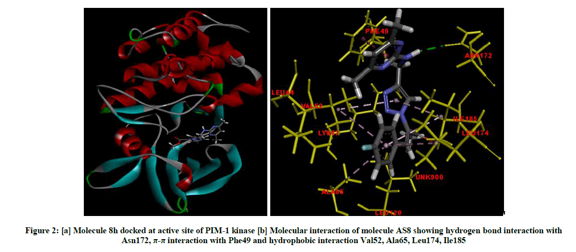
Figure 2: [a] Molecule 8h docked at active site of PIM-1 kinase [b] Molecular interaction of molecule AS8 showing hydrogen bond interaction with Asn172, π-π interaction with Phe49 and hydrophobic interaction Val52, Ala65, Leu174, Ile185
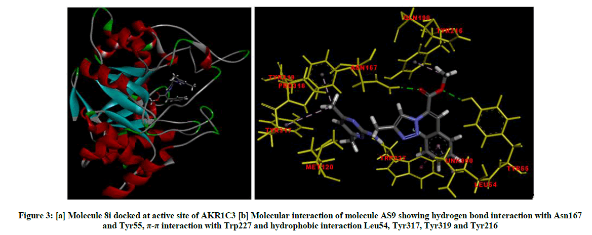
Figure 3: [a] Molecule 8i docked at active site of AKR1C3 [b] Molecular interaction of molecule AS9 showing hydrogen bond interaction with Asn167 and Tyr55, π-π interaction with Trp227 and hydrophobic interaction Leu54, Tyr317, Tyr319 and Tyr216
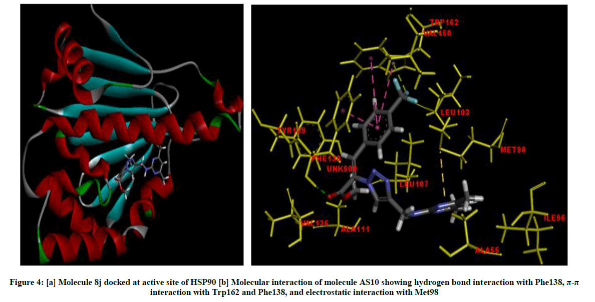
Figure 4: [a] Molecule 8j docked at active site of HSP90 [b] Molecular interaction of molecule AS10 showing hydrogen bond interaction with Phe138, π-π interaction with Trp162 and Phe138, and electrostatic interaction with Met98
ADME properties
The ADME properties (required pharmacokinetic properties of viable drug compounds) were calculated by Qikprop and analyzed by applying Lipinski's rule of five (Table 4) [44]. Molecular weight less than 650, partition coefficient between octanol and water (log Po/w) between −1 and 6.5 and solubility (log S) greater than -7. Pmdck greater than 5 and logBB greater than -3, this parameter tells us about the ability of the drug to pass through blood brain barrier. Majority of the molecules have the properties in range, partition coefficient of molecules ranged within the acceptable limits, solubility were in range except for molecule 8j that had values less than -7.0. Cell permeability and Blood Brain Barrier permeability for all molecules is in permissible range. Water solubility and oral absorption for half of the molecules is 100%. The values invariably imply that these molecules are potential drug molecules, which show better drug like property than standard doxorubucin and can be further optimized.
| Molecule | QP log Po/wa | QPlogSb | QPlogBBc | QPPMDCKd | Percent Human |
|---|---|---|---|---|---|
| Oral Absorptione | |||||
| 8a | 4.042 | -6.043 | -1.104 | 413.472 | 100 |
| 8b | 4.262 | -6.658 | -1.093 | 806.742 | 100 |
| 8c | 4.232 | -6.25 | -0.97 | 839.299 | 100 |
| 8d | 4.13 | -6.197 | -1.036 | 655.329 | 100 |
| 8e | 4.576 | -6.784 | -0.877 | 1465.139 | 100 |
| 8f | 4.247 | -6.08 | -0.922 | 856.135 | 100 |
| 8g | 3.174 | -5.238 | -1.759 | 99.734 | 74.747 |
| 8h | 4.004 | -6.282 | -1.138 | 591.231 | 100 |
| 8i | 3.904 | -6.031 | -1.149 | 460.105 | 100 |
| 8j | 4.743 | -7.333 | -1.005 | 1426.621 | 100 |
| Doxorubucin | -0.533 | -1.947 | -2.889 | 1.06 | 0 |
aPredicted octanol/water partition coefficient log P (acceptable range: 2.0-6.5);
bPredicted aqueous solubility in mol/l (acceptable range: 6.5-0.5);
cPredicted blood brain barrier permeability (acceptable range: 3-1.2);
dPredicted apparent MDCK cell permeability in nm/s (acceptable range in nm/s (acceptable range: <25 is poor and >500 is great);
ePercentage of human oral absorption (acceptable range: <25 is poor and >80% is high
Table 4: Calculated ADME properties of synthesized molecules
Molecular modeling studies
To understand the mode of binding and molecular interactions of 1,2,3-triazole derivatives, molecular docking studies were performed (Table 5). Crystal Structure of proteins were downloaded from protein data bank (PDB id: 1N37, 4AG8, 1WKQ, 5CE1, 5CF0, 5V82, 2YLP, 3SWZ, 4DBS) [45-51]. Interactions of the molecules with the proteins were analyzed to identify their hypothetical binding mode and binding affinity. All the molecular modeling calculations were performed using Schrödinger Suite 2010 on Linux platform. The proteins were prepared using protein preparation module applying the default parameters, hydrogen atoms were added and unwanted water molecules were removed from the protein structure followed by hydrogen bond optimization and energy minimization. Grid was generated around the active site (ligand binding site) defined by the co-crystallized ligand. Receptor Vander Waals scaling for the nonpolar atoms was set to 0.9 [52]. Molecules were built using Maestro build panel and prepared by LigPrep module using OPLS_2005 force field, GLIDE 5.6 was used for molecular docking. Low energy conformation of the ligands was selected and docked into the grid using extra precision (XP) docking mode. Further the absorption, distribution, metabolism and excretion (ADME) properties were calculated using Qikprop module in Schrodinger Suite.
| S. No. | Dock score in kcal/mol | ||||||||||
|---|---|---|---|---|---|---|---|---|---|---|---|
| 1N37 | 4AG8 | 4WKQ | 4ALW | 5CE1 | 5CF0 | 5V82 | 2YLP | 3SWZ | 4DBS | 5IRQ | |
| 1 | -3.831 | -7.804 | -5.63 | -6.26 | -6.518 | -9.836 | -4.624 | -3.966 | -6.672 | -8.553 | -6.275 |
| 2 | -5.008 | -7.575 | -6.419 | -5.76 | -6.514 | -8.094 | -8.952 | -3.867 | -6.4 | -8.878 | -6.944 |
| 3 | -4.396 | -7.871 | -5.494 | -5.898 | -6.453 | -9.103 | -7.413 | -5.373 | -7.47 | -8.635 | -6.792 |
| 4 | -3.946 | -7.829 | -4.972 | -4.942 | -6.477 | -9.659 | -9.312 | -4.14 | -6.724 | -8.488 | -6.494 |
| 5 | -3.326 | -6.616 | -5.396 | -4.724 | -5.763 | -8.587 | -4.483 | -3.22 | -7.431 | -8.32 | -6.549 |
| 6 | -2.941 | -8.092 | -6.651 | -5.726 | -7.249 | -9.798 | -9.212 | -4.263 | -8.532 | -8.522 | -6.83 |
| 7 | -4.329 | -6.298 | -5.183 | -5.411 | -5.982 | -8.732 | -8.735 | -2.617 | -6.163 | -7.792 | -5.075 |
| 8 | -4.612 | -6.958 | -5.622 | -6.136 | -5.958 | -9.11 | -9.568 | -4.952 | -5.495 | -8.16 | -6.313 |
| 9 | -4.211 | -7.81 | -6.721 | -5.66 | -5.719 | -9.491 | -9.11 | -6.108 | -7.495 | -9.206 | -6.322 |
| 10 | -5.075 | -7.902 | -6.151 | -8.872 | -5.565 | -9.71 | -8.053 | -3.492 | -6.414 | -8.748 | -4.648 |
| DOX | -11.258 | -12.456 | -14.423 | -10.315 | -7.396 | -7.842 | -12.82 | -11.34 | ---- | -12.246 | -7.719 |
Table 5: Dock score of molecules in all the protein used in docking studies
Conclusion
In conclusion, we demonstrated an efficient synthesis of a series of some novel Baylis-Hillman derived 1,2,3-triazole derivatives. The structures of newly synthesized compounds were characterized by IR, 1H-NMR, 13C-NMR and mass spectral analysis and also were screened for in vitro anticancer activity against six cancer cell lines and promising results are obtained. Based on the screening results, it was observed that three compounds 8h, 8i and 8j are proved to be most potent to anticancer activity against prostate cancer cell line (DU145) compared with reference to standard drug Doxorubicin. Additionally, molecular docking studies were carried out on DNA to predict the possible molecular target for the synthesised molecule and the values invariably imply that these molecules are potential drug candidates. Synthesis of nitrile containing Baylis Hillman target compounds and its library with further biological activity of these compounds is being studied and will be reported in due course.
Acknowledgement
The AS thanks the Principal, GDC (W) Hussainialam, for support and encouragement, UGC for sponsorship and CSIR-Indian Institute of Chemical Technology, for the facilities and encouragement. SS and CHNSSP thank VFSTR for encouragement. As acknowledge the UGC for sponsorship and sanction of project No. F. MRP-5717/15 (UGC-SERO).
References
- S.A. Rosenthal, H.M. Sandler, Nat. Rev. Urol., 2010,7, 31-38.
- H.H. Cheng, D.W. Lin, E.Y. Yu, Urol. Clin. North Am., 2012, 39, 561-571.
- T. Karantanos, P.G. Corn, T.C. Thompson, Oncogene., 2013, 32, 5501-5511.
- M.M. Centenera, A.K Fitzpatrick, W.D. Tilley, L.M. Butler, Biochim. Biophys. Acta., 2013,1835, 211-218.
- J. Ischia, F. Saad, M. Gleave, Curr. Opin. Urol., 2013, 23, 194-200.
- S. He, C. Zhang, A.A. Shafi, M. Sequeira, J. Acquaviva, J.C. Friedland, J. Sang, D.L. Smith, N.L. Weigel, Y. Wada, D.A. Proia, Int. J. Oncol., 2013, 42, 35-43.
- P. Martins, J. Jesus, S. Santos, L.R. Raposo, C. Roma-Rodrigues, P.V. Baptista, A.R. Fernandes, Molecules., 2015, 20, 16852-16891.
- H.C. Kolb, M.G. Finn, K.B. Sharpless, Angew. Chem. Int. Ed., 2001, 40, 2004-2021.
- E. Bokor, T. Docsa, P. Gergely, L. Somsak, Bioorg. Med. Chem., 2010, 18, 1171-1180.
- C. Gill, G. Jadhav, M. Shaikh, R. Kale, A. Ghawalkar, D. Nagargoje, M. Shiradkar, Bioorg. Med. Chem. Lett., 2008, 18, 6244-6247.
- R.P. Tripathi, A.K. Yadav, A. Ajay, S.S. Bisht, V. Chaturvedi, S.K. Sinha, Eur. J. Med. Chem., 2010,45, 142-148.
- S. Syed, M.M. Alam, N. Mulakayala, C. Mulakayala, G. Vanaja, A.M. Kalle, R. Reddanna Pallu, M.S. Alam, Eur. J. Med. Chem., 2012, 49, 324-333.
- B.S. Holla, M. Mahalinga, M.S. Karthikeyan, B. Poojary, P.M. Akberali, N.S. Kumari, Eur. J. Med. Chem., 2005, 40, 1173-1178.
- K.D. Thomas, A.V. Adhikari, N. S. Shetty, Eur. J. Med. Chem., 2010, 45, 3803-3810.
- N.G. Aher, V.S. Pore, N.N. Mishra, A. Kumar, P.K. Shukla, A. Sharma, M.K. Bhat, Bioorg. Med. Chem. Lett., 2009, 19, 759-763.
- J.N. Sangshetti, R.R. Nagawade, D.B. Shinde, Bioorg. Med. Chem. Lett., 2009, 19, 3564-3567.
- J.N. Sangshetti, D.B. Shinde, Bioorg. Med. Chem. Lett., 2010,20, 742-745.
- L. Zhou, A. Adel, M. Korn, R. Burda, J. Balzarini, E. De Clercq, E.R. Kern, P.F. Torrence, Antivir. Chem. Chemother., 2005, 16, 375-383.
- A.S. El-Etrawy, A.A. -H. Abdel-Rahman, Chem. Heterocycl. Compd., 2010, 46, 1105-1108.
- W.Q. Fan, A.R. Katritzky, in A.R. Katritzky, C.W. Rees, E.F.V. Scriven, (Eds), Comprehensive Heterocyclic Chemistry II, Vol. 4, Elsevier Science: Oxford, 1996, pp 1-126.
- D.R. Buckle, C.J. Rockell, H. Smith, B.A. Spicer, J. Med. Chem., 1986, 29, 2262-2267.
- M.H. Taylor, A. Sandler, W.J. Urba, A.M.P. Omuro, G.S. Gorman, R.A.Karmali, J. Cancer Ther., 2015, 6, 322-333.
- S.E. Drewes, G.H.P. Roos, Tetrahedron., 1988, 44, 4653-4670.
- D. Basavaiah, P. Dharma Rao, R. Suguna Hyma, Tetrahedron., 1996, 52, 8001-8062.
- E. Ciganek, Organic Reactions; L. A., Ed.; Wiley: New York, 1997; Vol. 51, p 201.
- V. Singh, S. Batra, Tetrahedron., 2008, 64, 4511-4574.
- G.N. Ma, J.J. Jiang, M. Shi, Y. Wei, Chem. Commun., 2009, 5496-5514.
- A. Santhoshi, B. Mahendar, S. Mattapally, P.S. Sadhu, S.K. Banerjee, V. Jayathirtha Rao, Bioorg. Med. Chem. Lett., 2014,24, 1952-1957.
- Ch.N.S.S.P. Kumar, D.K. Parida, A. Santhoshi, A.K. Kota, B. Sridhar, V. Jayathirtha Rao, Med. Chem. Commun.,2011,2, 486-492.
- A. Santhoshi, P.S. Sadhu, R. Sriram, Ch.N.S.S.P. Kumar, B. Mahendar, V. Jayathirtha Rao, Med. Chem. Res., 2013, 22, 3329-3340.
- S. Bharat Kumar, Ch.N.S.S.P. Kumar, A. Santhoshi, K. Pranay Kumar, U.S.N. Murthy, V. Jayathirtha Rao, Synth. Commun., 2017, 47, 131-136.
- J. Cai, Z. Zhou, G. Zhao, C. Tang, Org. Lett., 2002, 4, 4723-4725.
- D.J. Maher, S.J. Connon, Tetrahedron Lett., 2004, 45, 1301-1305.
- D. Basavaiah, M. Krishnamacharyulu, R.S. Hyma, P.K.S. Sarma, N. Kumaragurubaran, J. Org. Chem., 1999, 64, 1197-1200.
- M. Sa Marcus, M.D. Ramos, L. Fernandes, Tetrahedron., 2006, 62, 11652-11656.
- V. Vichai, K. Kirtikara, Nature protocols., 2006, 1, 1112-1116.
- F.A. Fornari, J.K. Randolph, J.C. Yalowich, M.K. Ritke, D.A. Gewirtz, Mol. Pharmacol., 1994, 45, 649-656.
- T.J. Lynch, D.W. Bell, R. Sordella, S. Gurubhagavatula, R.A. Okimoto, B.W. Brannigan, P.L. Harris, S.M. Haserlat, J.G. Supko, F.G. Haluska, D.N. Louis, D.C. Christiani, , J. Settleman, D. A. Haber, N. Eng. J. Med., 2004, 350, 2129-2139.
- A. Sawai, S. Chandarlapaty, H. Greulich, M. Gonen, Q. Ye, C. L. Arteaga, W. Sellers, N. Rosen, D. B. Solit, Cancer Res., 2008, 68(2), 589-596.
- M. Bachmann, T. Möröy, Int. J. Biochem. Cell Biol., 2005, 37(4), 726-730.
- N.M. DeVore, E.E. Scott, Nature., 2012, 482(7383), 116-119.
- M.C. Byrns, Y. Jin, T.M. Penning, J. Steroid Biochem. Mol. Biol., 2011, 125, 95-104.
- F. Alimirah, J. Chen, Z. Basrawala, H. Xin, D. Choubey, FEBS Lett., 2006, 580(9), 2294-2300.
- C.A. Lipinski, F. Lombardo, B.W. Dominy, P.J. Feeney, Adv. Drug Deliv. Rev., 1997, 23, 3-25.
- M.S. Searle, A.J. Maynard, H.E.L. Williams, Org. Biomol. Chem., 2003, 1, 60-66.
- M. McTigue, B.W. Murray, J.H. Chen, Y.L. Deng, J. Solowiej, R.S. Kania, Proc. Natl. Acad. Sci. USA., 2012, 109, 18281-18289.
- ]S.H. Liaw, Y.J. Chang, C.T. Lai, H.C. Chang, G.G. Chang, J. Biol. Chem., 2004, 279, 35479-35485.
- J. Li, F. Shi, D.Q. Chen, H.L. Cao, , B. Xiong, J.K. Shen, J.H. He, Nucl. Sci. Tech., 2015, 26, 060502-060502.
- X. Wang, A. Kolesnikov, S. Tay, G. Chan, Q. Chao, S. Do, J. Drummond, A.J. Ebens, N. Liu, J. Ly, E. Harstad, H. Hu, J. Moffat, V. Munugalavadla, J. Murray, D. Slaga, V. Tsui, M. Volgraf, H. Wallweber, J.H. Chang, J. Med. Chem., 2017, 60, 4458-4473.
- N.A. Lack, P. Axerio-Cilies, P. Tavassoli, F.Q. Han, K.H. Chan, C. Feau, E. Leblanc, E.T. Guns, R.K. Guy, P.S. Rennie, A. Cherkasov, J. Med. Chem., 2011, 54, 8563-8573.
- M. Chen, A.O. Adeniji, B.M. Twenter, J.D. Winkler, D.W. Christianson, T.M Penning, Bioorg. Med. Chem. Lett., 2012, 22, 3492-3497.
- R.A. Friesner, J.L. Banks, R.B. Murphy, T.A. Halgren, J.J. Klicic, D.T. Mainz, M.P. Repasky, E.H. Knoll, M. Shelley, J.K. Perry, D.E. Shaw, P. Francis, P.S. Shenkin, J. Med. Chem., 2004, 47, 1739-1749.


