Research Article - Der Pharma Chemica ( 2018) Volume 10, Issue 2
In Vitro Enzymatic Assays of Azadirachta indica Extract and Identification of Bioactive Compounds using Gas Chromatography-Mass Spectrophotometer
Adewole E1,4*, Ojo A1, Omoaghe AO2 , Enye LA3 and Jamshed I4
1Department of Chemical Sciences, Afe Babalola University Ado, Ekiti State, Nigeria
2Department of Physiology, Afe Babalola University Ado, Ekiti State, Nigeria
3Department of Anatomy, Afe Babalola University Ado, Ekiti State, Nigeria
4Department of Pharmacy, Centre for Advanced Drug Research (CADR), COMSATS Institute of Information Technology, Abbottabad, Pakistan.
- *Corresponding Author:
- Adewole E
Department of Chemical Sciences
Afe Babalola University Ado
Ekiti State, Nigeria
Abstract
Background: Azadirachta indica leaves have been discovered to be a therapeutic plant with various pharmacological properties and are used locally for treating different types of ailments which include malarial, diabetes mellitus and a host of others. The plant after extensive literature search has not been simultaneously been investigated for its antidiabetic potentials against the α-glucosidase, β- glucosidase, maltase glucoamylase and the aldose reductase which has two isoforms (ALR1 and ALR2) and this necessitate the research work.
Methods: The in vitro enzymatic assays were done after the subjecting the plant to extraction using chloroform solvent. The percentage inhibition of α-glucosidase, β- glucosidase and maltase glucoamylase were determined and the aldose reductase which has two isoforms (ALR1 and ALR2) inhibitory potential and inhibitory concentration were determined using laid down protocol with slight modification and finally the crude extract was characterized using GC-MS to revealed the presence of bioactive compounds which were screened computationally for their various drug properties using OSIRIS Online Server Explorer.
Results: The extract percentage inhibition against α-glucosidase was 76.95+4.19%, maltase glucoamylase (73.9+5.92%), the results were better than the standard acarbose (68.54 ± 8.48), β-glucosidase (18.03 ± 4.59) showed less potent compared with standard castanospermine (59.98). The IC50 of ALR1 and ALR2 were in the same range of 1.05+0.5 μg/ml. The GC-MS revealed compounds such as D-mannitol,1-decysulfonyl, Z-27-Hexatriacontan-2-one, oxalic acid,6-ethyl oct-3-yl-isobutyl ester, Lanosta-7,9 (11)-dien- 18-oic Acid, 22,25-epoxy-3,17,20-trihydroxy-gamma-lactone, 1,3-dioxolane,4-ethyl-5-octyl,2,2-bis(trifluoro methyl)-, trans, pyrrolidine,1- methyl-3,2-spiro-benzo-1,3-dioxolane. 3-Tosyl sedoheptulose and carbonic acid, pentadecyl ester and these compounds exhibited various when screened computionall using Online OSIRIS explorer for their various drug properties.
Conclusion: The potent inhibitory activities of the chloroform extract against α-glucosidase, maltase glucoamylase and the aldose reductase (ALR1 and ALR2) is an indication that the plant possesses good therapeutic potential for the treatment against diabetes mellitus.
Keywords
Diabetes mellitus, Aldose reductase, α-glucosidase, Glucoamylase, β-Glucosidase, Bioactive compounds and drug properties
Introduction
Diabetes mellitus is said to a class for metabolic disorders associated with high blood sugar contents for a long period of time [1]. Medical symptoms of high blood sugar include constant urination, increased hunger and thirst, while acute complications include diabetic ketoacidosis, hyperosmolar hyperglycemic condition or death, moreover long term effect include damage to eyes, kidney problem, stroke, death cardiovascular disease [2]. The pharmacological mechanism of diabetes is either due to failure of pancreas unable to produce sufficient insulin or not responding to insulin produced [3]. There are many anti-diabetes inhibitors and among which are α-glucosidase, β-glucosidase. α- glucosidase inhibitors are oral drugs made for Diabetes Mellitus Type-2 (DMT-2) and it works by disallowing the digestion of carbohydrates and thereby reducing the impact of carbohydrate on blood sugar. Generally, plants are considered to be a source for the most active, potent hypoglycaemic properties [4,5]. Natural drugs from plants are considered to be non0toxic with lesser side effects than synthetic drugs. It has been reported that medicinal plants possessing anti-diabetic activities could be a useful tool for the discovery of safer hypoglycaemic agents [6]. These plants are said to be the major source for discovering new compounds with therapeutic value for drug development against most common and very prevalent disease, diabetes mellitus. The plants which have therapeutic application possess bioactive composites viz., alkaloids, glycosides, tannins, flavonoids, saponins, phenolics and vitamins [7]. It has been earlier reported that Aldose reductase having two isoforms (ALR1 and ALR2) has been implicated in the cause of diabetic problems that may be connected to a much significance flux of glucose through the polyol pathway, caused in tissues such as lens, kidney, retina and nerves at high blood glucose levels. Therefore, the inhibitory property of aldose reductase is gaining recognition as a major therapeutic tool for the treatment of hyperglycemia-induced cardiovascular pathologies [8]. Long term drawbacks of diabetes include among others; cataract genesis and microangiopathy (Including nephropathy, retinopathy and neuropathy) thought to be linked to excess free glucose in corresponding tissues [9]. Widely research work has provided evidence of Aldose Reductase (AR) implication in diabetes severalty [10]. Scientific investigation to provide inhibitors of AR has been a major task for the chemists in order to effectively treat various degrees of complications associated with diabetes.
Azadirachta indica is a plant having the following taxonomy: order: Rutales, suborder Rutinae, family: Meliaceae (Mahogany family), subfamily: Melioideae, tribe: Melieae, genus: Azadirachta, species indica [11]. All the parts of the plants have been research and found that they possess various pharmacological properties which are being employed in treatment of different ailments such as diabetes, cancer, fungicidal diseases. It has been reported that the aqueous leaves extract have been found to reduced considerably the blood sugar level and prevent adrenaline as well as glucose induced hyperglycaemia [12] Also, the aqueous leaf extract reduces hyperglycaemia in streptozotocin diabetes and the effect is possibly due to presence of a flavonoid, quercetin as reported by Chakraborty [13]. The mechanisms underlying the hypoglycaemic of the aqueous leaf extract have also been discussed as reported by [14,15]. However, chloroform extracts have not been concurrently being examined for their inhibitory potentials against α, β-glucosidases, maltase glucoamylase and aldose reductase (ALR1 and ALR2).
Method
Identification
After extensive literature search, it was discovered that plant has not been evaluated concurrently for its potent inhibition of α- and β- glucosidase, maltase glucoamylase, aldose reductase (ALR1 and ALR2) and identifying the bioactive compounds of the chloroform crude extract.
Methodology
Plants source
A. indica, leaves were collected from farm and were authenticated at the Department of Agricultural Science, Afe Babalola University Ado Ekiti, Nigeria.
Crude extracts preparation
The extraction was done by soaking two hundred grams of powdered samples in 1000 ml of chloroform for a period of 5 days and filtered through Whatman filter paper. The extracts were concentrated using a rotary evaporator at 35°C and the dried extract was stored at room temperature for further use. The stock sample was prepared by dissolving 10 mg of dried crude extracts in 1 ml of 100% Dimethyl Sulfoxide (DMSO) and labeled as stock (10 mg/ml), working solution was made as 1 mg/ml.
Maltase-glucoamylase inhibition assay
Maltase-glucoamylase inhibition assay was carried out using the substrate p-nitrophenyl α-d-glucoside using reported procedure [16]. The reaction mixture contained 70 μl buffer, 10 μl extracted enzyme (25.0 μg of protein) and 10 μl of test compounds and incubated at 37°C for 5 min. After the incubation, 10 μl of p-NPG (10 mM, prepared in assay buffer) was added to each well of a 96-well plate and further incubated at 37°C for 30 min. The activity of the test compounds against maltase glucoamylase was determined by measuring p-nitrophenol at a wavelength of 405 nm. Acarbose was used as a positive control. The percent inhibition was calculated using the following equation:
α-glucosidase inhibition study
The inhibitory effect of the 1 mg/ml of the working solution was performed by slight modification of a previously published method [17]. Briefly, solutions of α-glucosidase (from Saccharomyces cerevisiae) and its substrate p-nitrophenyl α-D-glucopyranoside (pNPG) were prepared in phosphate buffer (70 mM, pH 6.8). Buffer was used for the preparation of inhibitor solutions. The inhibition assays were conducted by adding inhibitor solution (10 μl) to 70 μl buffer and 10 μl of enzyme solution (2.5 unit/ml) in 70 mM phosphate buffer (pH 6.8) followed by pre-incubation at 37°C for 5 min. After pre-incubation, 10 μl of 10 mM substrate (pNPG) prepared in phosphate buffer was added to the mixture to initiate enzymatic reaction. The reaction mixture was incubated at 37°C for 30 min. Acarbose was used as a positive control. The α-glucosidase activity was determined by measuring the p-nitrophenol released from pNPG at 405 nm using an Elx 800 Micro plate reader.
β-glucosidase inhibition study
The evaluation of inhibitory activity against β-glucosidase was performed with slight modification of the previously published method [18]. Briefly, β-glucosidase (From sweet almonds) enzyme and p-nitrophenyl β-D-glucopyranoside (pNPG) as substrate were prepared in 0.07 M phosphate buffer (pH 6.8). The inhibition assays were conducted by adding inhibitor solution (10 μl) to 70 μl buffer and 10 μl of enzyme solution (2.0 unit/ml) in 0.07 M phosphate buffer (pH 6.8) followed by pre-incubation at 37°C for 5 min. Following pre-incubation, 10 μl of 10 mM p-nitrophenyl Glucopyranoside (pNPG) in phosphate buffer was added as a substrate to the mixture to start the reaction. The reaction mixture was then incubated at 37°C for 30 min. Negative control contained 10 μl of 10% DMSO instead of inhibitor. Castanospermine was used as a positive control.
Determination of Aldose Reductase (ALR2) inhibitory activity
UV spectrophotometer was used at 340 nm in order to determine the activity of aldose reductase by measuring the Nicotinamide Adenine Dinucleotide Phosphate (NADPH) consumption. Each well of the 96-well plate contains exactly 100 μl of assay mixture containing phosphate buffer 100 mM at pH 6.2 (10 μl), with 10 μl of 1 mg/ml of crude extracts followed by addition of 35 μl of enzyme and 20 μl of substrate (D,L-glyceraldehyde). The mixture was incubated at 37°C for 5 min and for the enzymatic reaction to run properly 0.5 mM NADPH (20 μl) as a cofactor was added and reading was taken at 340 nm. The mixture was incubated again at 37°C for 10 min and reading was taken at the respective UV range in Enzyme-linked Immunosorbent Assay (ELISA) plate reader. As positive and negative control, 10 μl of 10 mM Sorbini and 20 μl buffer solution, respectively, were used. The enzymatic reaction was run in triplicate with a final volume of 100 μl in each well. Absorbance was noted and results were analysed.
Determination of Aldehyde Reductase (ALR1) inhibitory activity
UV spectrophotometer was used at 340 nm in order to determine the activity of aldehyde reductase by measuring the NADPH consumption. Each well of the 96-well plate contains exactly 100 μl of assay mixture containing phosphate buffer 100 mM at pH 7.2 (10 μl), with 10 μl of 1 mg/ml of crude extracts followed by addition of 35 μl of enzyme and 20 μl of substrate (sodium D-glucoronate). The mixture was incubated at 37°C for 5 min and for the enzymatic reaction to run properly 0.5 mM NADPH (20 μl) as a cofactor was added and reading was taken at 340 nm. The mixture was incubated again at 37°C for 5 min and reading was taken at the respective UV range in ELIZA plate reader. As positive and negative control, 10 μl of 10 mM vaproic acid and 20 μl buffer solution, respectively, were used. The enzymatic reaction was run in triplicate with a final volume of 100 μl in each well. Absorbance was noted and results were analysed.
GC/MS analysis
GC-MS analysis of methanol and chloroform extracts of were performed using TurboMass GC System, fitted with an Elite-5 capillary column (30 m, 0.25 mm inner diameter, 0.25 μm film thickness; maximum temperature, 350°C coupled to a Perkin Elmer Clarus 600C MS. Helium was used as gas carrier at a constant flow rate of 1.0 ml/min. The injection, transfer line and ion source temperatures were 280°C. The ionizing energy was 70 eV. The oven temperature was programmed from 70°C (hold for 2 min) to 280°C (hold for 10 min) at a rate of 5°C/min. The crude extract was solubilized with chloroform and filtered with syringe filter (Corning, 0.45 μm). Volumes of 1 μl of the crude extracts were injected with a split ratio 1:20. The data were obtained by collecting the mass spectra within the scan range 50-550 m/z. The identification of chemical compounds in the extract was based on GC retention time; the mass spectra matched those of standards available at NIST library.
Statistical analysis
The inhibition analysis was performed in triplicates and the total percentage inhibitions were calculated by method of (Tables 2 and 3) [17]:
ALR1 and ALR2 IC50 (i.e., the concentration of sample inhibiting 50%) values of potent inhibitors were determined by testing the serial dilutions of inhibitors and were calculated by using the program PRISM 5.0 (GraphPad, San Diego, California, USA).
Discussion
From the result of Table 1 and Figure 1, the chloroform extract of A. indica had 76.95 ± 4.19% against α-glucosidase and the result was better than the standard acarbose having 68.54 ± 8.48%. Also, the maltase glucosidase inhibitory potential of chloroform extract was higher (76.91 ± 5.92% inhibition) than the acarbose, however, the extract demonstrated very poor potent inhibition against beta glucosidase (18.03 ± 4.59% inhibition) compared with the castanospermine standard (59.98%). These ALR2 value of the chloroform extract (IC50 1.047 ± 0.50 μg/ml) showed very promising selective inhibitor activities when compared with the standard positive control, sorbinil of IC50 (3.10 ± 0.20 μM). The Aldehyde Reductase (ALR1) value IC50 (1.050 ± 0.50 μg/ml) was better when compared to standard 10 mM vaproic acid IC50 (57.4 ± 10 μM).
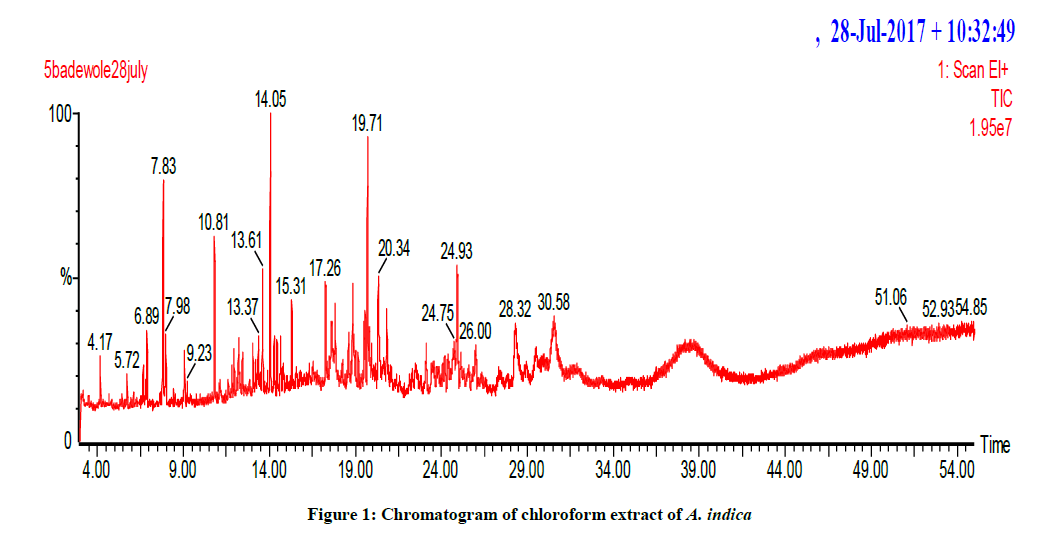
Figure 1: Chromatogram of chloroform extract of A. indica
| Extract | α-glucosidase (% inhibition) |
Maltase glucoamylase (% inhibition) |
β-glucosidase (% inhibition) |
ALR1 IC50 (μg/ml) |
ALR2 IC50 (μg/ml) |
|---|---|---|---|---|---|
| A. indica | 76.95 ± 4.19 | 73.91 ± 5.92 | 18.03 ± 4.59 | 1.050 ± 0.5 | 1.047 ± 0.5 |
| Acarbosea | 68.54 ± 8.48 | 68.54 ± 8.48 | Not tested | Not tested | Not tested |
| Castanospermineb | Not tested | Not tested | 59.98 [19,20] | Not tested | Not tested |
| Vaproic acidc | Not tested | Not tested | Not tested | 57.4 +10 | Not tested |
| Sorbinild | Not tested | Not tested | Not tested | Not tested | 3.10 ± 0.20 |
SEM ± Standard mean error; aα-glucosidase and maltase glucoamylase standards; bβ-glucosidase standard; cALR1 standard; dALR2 standard (dReported IC50 of 3.42 μM of Sorbinil by Rakowitz et al. 17a and Ali et al.17b)
Table 1: Inhibitory effect of chloroform crude extract
| Compound name | Molecular formula | Molecular weight | CAS No | Retention time (min) |
|---|---|---|---|---|
| D-mannitol,1-decysulfonyl | C16H34O7S | 370 | 900154-76-1 | 49.928 |
| Pyrrolidine,1-methyl-3,2-spiro-benzo-1,3-dioxolane | C11H13O2N | 191 | 40259-14-1 | 20.296 |
| 3-Tosyl sedoheptulose | C14H20O9S | 364 | 900126-65-3 | 32.435 |
| Phenol,3,5-bis(1,1-dimethyl) | C14H22O | 206 | 1138-52-9 | 20.346 |
| Carbonic acid, butyl pentadecyl ester | C20H40O3 | 328 | 900314-64-2 | 43.010 |
| 1,3-Dioxolane,4-ethyl-5-octyl,2,2-bis(trifluoro methyl)-, trans | C15H24O2F6 | 350 | 38274-73-6 | 44.511 |
| Lanosta-7,9 (11)-dien-18-oic acid, 22,25-epoxy-3,17,20-trihydroxy-gamma-lactone | C30H44O5 | 484 | 56143-25-0 | 29.285 |
| Oxalic acid,6-ethyl oct-3-yl-isobutyl ester | C16H30O4 | 286 | 900309-34-1 | 24.958 |
| Z-27-Hexatriacontan-2-one | C36H70O | 518 | 130385-25-0 | 40.879 |
Table 2: Some identified compounds in the crude extract of A. indica
| Compound | Drug likeness | Mutagenic | Tumorigenic | cLogS | cLogP | Polar surface area (A°) |
%Absorption | H-bond Acceptor | H-bond Donor | Reproductivity | Sp3 atoms | Aromatic ring |
|---|---|---|---|---|---|---|---|---|---|---|---|---|
| aMannitol | -0.1995 | None | None | 0.546 | -3.1458 | 0.60874 | 108.79 | 6 | 6 | none | 12 | 0 |
| bCarbonic Acid | -2.521 | None | High | -0.646 | -0.5238 | 0.89283 | 108.69 | 3 | 2 | none | 2 | 0 |
| cPhenol | -2.2721 | High | High | -1.32 | 1.3139 | 0.16455 | 108.94 | 1 | 1 | high | 1 | 0 |
| dOxalic acid | -6.1289 | High | None | 0.066 | -1.5754 | 0.84733 | 108.71 | 4 | 2 | high | 2 | 0 |
| eSedoheptulose | 1.9967 | None | None | 0.624 | -3.7444 | 0.62395 | 108.78 | 7 | 6 | high | 12 | 0 |
| fPyrrolidine | -0.72478 | High | High | -0.855 | 0.4025 | 0.17233 | 108.94 | 1 | 1 | high | 5 | 0 |
| gGamma-Butyrolactone | -5.6615 | High | High | -0.973 | -0.0202 | 0.33377 | 108.88 | 2 | 0 | high | 4 | 0 |
| hDioxolane | -6.9824 | High | None | -0.901 | -0.1434 | 0.32637 | 108.88 | 2 | 0 | high | 5 | 0 |
aMannitol: Shares the same functional group with D-mannitol,1-decysulfonyl;
bCarbonic acid: Shares the same functional group with carbonic acid, butyl pentadecyl ester;
cphenol: Shares the same functional group with Phenol,3,5-bis(1,1-dimethyl);
dOxalic acid: Shares the same functional group with oxalic acid,6- ethyl oct-3-yl-isobutyl ester;
esedoheptulose: shares the same functional group with 3-Tosyl sedoheptulose;
fPyrrolidine: Shares the same functional group with pyrrolidine,1-methyl-3,2-spiro-benzo-1,3-dioxolane;
gGamma-butyrolactone: shares the same functional group with lanosta-7,9 (11)-dien-18-oic acid,22,25-epoxy- 3,17,20-trihydroxy-gamma-lactone;
hDioxolane shares the same functional group with 1,3-dioxolane,4-ethyl-5-octyl,2,2-bis(trifluoro methyl)-trans
Table 3: Drug properties of some of identified compounds in the crude extracts determined by OSIRIS property explorer
It has been severally reported that the antidiabetic potentials of plants are connected to the presence of phytochemicals and these phytochemical includes among others flavonoids, saponin, phenolic compounds, alkaloids, glycosides and are highly potent hypoglycaemic agents. The aqueous and ethanolic extracts of A. indica has been previously evaluated for the presence of phytochemicals as reported by Prashanth [20], qualitative evaluation has shown the presence of alkaloids, carbohydrates, reducing sugars, flavonoids, glycosides, tannins and phenolic compounds, triterpenoids and steroids. It has been documented that inhibition of α-glucosidase and α-amylase activity results in slowing down the carbohydrate digestion of absorbable monosaccharide’s, causing reduction of postprandial hyperglycaemia. The plant extract showed high inhibition against α-glucosidase (76.95 ± 4.19%), maltase glucoamylase of (73.91 ± 5.92%) when compared to the standard acarbose (68.54 ± 8.48%). α-glucosidase inhibitors delay intestinal carbohydrate absorption and slow the sharp rise in blood sugar levels that diabetic patients typically experience after taken food very rich in sugar content. In contrast, none of the currently available α-glucosidase inhibitors for clinical use are without severe side effects [21-23]. The searches for new therapeutic agents from medicinal plants have therefore; become an attractive approach for the treatment of postprandial hyperglycemias.
The presence of various bioactive compounds as revealed by the Gas Chromatography-Mass Spectrophotometer (GC-MS) is an indication that the plant, A. indica has highly potent therapeutic property as antidiabetes agent, due to the presence of compounds identified in the chloroform extract and these include pyrrolidine,1-methyl-3,2-spiro-benzo-1,3-dioxolane, carbonic acid, butyl pentadecyl ester, d-mannitol,1-decysulfonyl, phenol,3,5-bis(1,1-dimethyl), lanosta-7,9 (11)-dien-18-oic acid, 22,25-epoxy-3,17,20-trihydroxy-gamma-lactone and 1,3-dioxolane,4-ethyl-5- octyl,2,2-bis(trifluoro methyl)-, trans. these heterocyclic compounds have been found to possess various pharmacological activities such as antidiabetic, antifungal and host of others. It has been reported by Ana Trapero [24] that antidiabetic α-glucosidase inhibitors share some common structural characteristics with a class of bioactive molecules that mimic the sugar structures and generally, these are cyclic compounds containing a basic amine functionality with several hydroxyl substituents that have a tridimensional arrangement similar to the present in elemental carbohydrates [24] and these structural similarities are translated to the carbohydrate functional roles, and numerous members of these families display interesting biological and enzymatic activities, especially as glycosidase inhibitors [24]. In addition to for the search of drug properties of the identified compounds, The online OSIRIS property explorer server has revealed the relevance and various drug properties of some of the identified compounds in the crude extracts using GC-MS and this could serve as a tool for the pharmaceutical chemists to further research on the plant as a potential anti-diabetic agent.
Statement of significance
This study discovered the potent anti-diabetic inhibitory potentials of the plant against α-glucosidase and maltase glucoamylase which can be highly beneficial for the treatment of diabetes mellitus type 2. Also, the online OSIRIS server explorer has revealed the drug properties of some of the identified compounds which will be beneficial to the scientists for further investigation. This study could be explored by researchers and a new anti-diabetic agent may be arrived at.
Acknowledgement
The author sincerely appreciates ‘The World Academy of Science’ (TWAS) for the Postdoctoral fellowship opportunity granted at the Centre for Advanced Drug Research, COMSATS Institute of Information Technology, Abbottabad, Pakistan in 2017.
References
- About Diabetes, World Health Organization, Archived from the original on 31 March, 2014.
- Diabetes Fact Sheet No 312", WHO. October 2013, Archived from the original on 26 August, 2013.
- Shoback, edited by David G. Gardner, Dolores, Chapter 17". Greenspan's Basic & Clinical Endocrinology, 9th Edi., New York: McGraw-Hill Medical., 2011.
- D.K. Patel, R. Kumar, D. Laloo, S. Hemalatha, Asian Pac. J. Trop. Dis.,2012a, 2, 239-250.
- D.K. Patel, S.K. Prasad, R. Kumar, S. Hemalatha, Asian Pac. J. Trop. Biomed., 2012b, 2, 320-330.
- C. Sunila, P. Agastian, C. Kumarappan, S. Ignacimuthu, Food Chem. Toxicol.,2012, 50, 1547-1553.
- A. Ghani, Medicinal Plants of Bangladesh, 2nd Edi., 2003, 1-2, 55-57, 402, 500.
- S.M. Grundy, I.J. Benjamin, G.L. Burke, A. Chait, R.H. Eckel, B.V. Howard, W. Mitch, S.C. Jr. Smith, J.R. Sowers, Circulation.,1999, 100, 1134-1146.
- P. Fresneau, M. Cussac, J.M. Morand, B. Szymonski, D. Tranqui, G. Leclerc, J. Med. Chem., 1998, 41(24), 4706-4715.
- D.R. Tomlinson, G.B. Willars, A.L. Carrington, Pharmacol. Ther., 1992, 54(2), 151-199.
- K. Biswas, I. Chattopadhyay, K. Ranajit, Curr. Sci., 2002, 82, 11.
- K.S. Murty, D.N. Rao, D.K. Rao, L.B.G. Murty, Indian J. Pharmacol., 1978, 10, 247-250.
- T. Chakraborty, L. Uerotta, G. Poddar, Phytother. Res., 1989, 3, 30-32.
- R.R. Chattopadhyay, Gen. Pharmacol., 1996, 27, 431-434.
- R.R. Chattopadhyay, J. Ethnopharmacol., 1999, 67, 373-376.
- A. Tanaka, M. Ohya, T. Yammato, C. Nakagawa, T. Tsuji, K. Senoo, H. Obata, Biosci. Biotechnol. Biochem.,1999, 63, 1548-1552.
- H.Y. Ma, H.Y. Gao, L. Sun, J. Huang, X.M. Xu, L.J. Wu, J. Nat. Med., 2011, 65, 37-42.
- M. Pérez, F.J. Muñoz, E. Muñoz, M. Fernández, J.V. Sinisterra, M.J. Hernáiz, J. Mol. Catal. B: Enzym., 2008, 52-53, 153-157.
- D. Rakowitz, R. Maccari, R. Ottana, M.G. Vigorita, Bioorg. Med. Chem., 2006, 14, 567-574.
- S. Ali, A. Saeed, N. Abbas, M. Shahid, M. Bolte, J. Iqbal, Med. Chem. Commun., 2012. 2012, 3, 1428-1434.
- K. Shwetha, H.N. Narasimha Murthy, K.V.S. Rajeswara Rao, N.S. Narahari, R. Agarwal, R. Singh, IJAETMAS., 2017, 5(1).
- H. Kurihara, J. Ando, M. Hatano, J. Kawabata, Bioorg. Med. Chem. Lett., 2011, 5, 1241-1244.
- K. Sama, K. Murugesan, R. Sivaraj, Asian J. Plant Sci. Res., 2012, 4, 550-553.
- A. Trapero, A. Llebaria, J. Med. Chem., 2012, 55, 10345-10346.


