Research Article - Der Pharma Chemica ( 2018) Volume 10, Issue 5
Isolation and Characterization of Antibacterial Endophytic Bacteria from Mangrove Plants at Kapo-Kapo and Setan Islands, West Sumatra Indonesia
Anthoni Agustien1*, Akmal Djamaan2, Yetria Rilda3 and Nasril Nasir1
1Department of Biology, Andalas University, Kampus Limau Manis, Padang, Indonesia
2Faculty of Pharmacy, Andalas University, Kampus Limau, Padang, Indonesia
3Department of Chemistry, Andalas University, Kampus Limau Manis, Padang, Indonesia
- *Corresponding Author:
- Anthoni Agustien
Department of Biology
Andalas University
Kampus Limau Manis, Padang, Indonesia
Abstract
Endophytic bacteria is a group of microorganisms that have an important role in secondary metabolite income. Leaf sampling from mangrove plants was done purposive random sampling. Isolation of endophytic bacteria with spread plate method. Screening of endophytics for antibactrial producing is done by paper disc method. Five antibacterial bacterial isolates were obtained on mangrove leaf, three isolates had inhibition zone against Escherchia coli bacteria, four isolates had inhibition zone against Staphylococcus aureus bacteria and two bacterial isolates against both test bacteria. Physiological characterization and biochemical test were obtained for four suspected species, Enterobacter sp., Bacillus sp.1, Klebsiella sp. and Bacillus sp.2.
Keywords
Isolation, Characterization, Endophytic bacteria, Antibacterial, Mangrove
Introduction
Endophytes isolated from mangrove plants are highly potential for the livelihood of new natural products. The unique mangrove plant environment of semi-salinity provides an opportunity for microorganisms as endophytic microbes are widespread in leaves and other organs of host plants [1]. Mangrove plants have historically explored intensively for health purposes, mangrove leaf extract has antibacterial activity against pathogenic bacteria and Candida spp. [2]. Many of endophytes are capable of synthesizing bioactive compound that can be used by plants for defense against pathogens and some of these compounds have been proven useful for drug discovery [3].
Indonesia is a country that has high biodiversity and possesses tropical rain forest area so it is an advantage in the sourcing of bioactive compounds [4]. West Sumatra has mangrove forests in several places or locations on the coast. Research on mangrove analysis in Pesisir Selatan district, obtained 15 species with 658 individual mangroves. The dominant mangrove plant in Kapo-kapo is Rhizopora apiculata, Rhizopora sp., Sonneratia alba and Lumnitzera littorea whereas on the island of Setan, Rhizopora apiculata, Rhizopora are dominating. The aim of this research is to isolation of antibacterial endophytic bacteria from mangrove plants endophytic bacteria isolate mangrove plant and characterize endophytic bacterial isolates that are potential anti-bacterial producers.
Materials and Methods
Collection of leaf samples from mangrove plants
Leaf sampling from Rhizopora apiculata, Rhizopora sp.1, Sonneratia alba, Lumnitzera littorea mangrove plants was done by purposive random sampling method from Kapo-kapo and Setan islands area location, district of Pesisir Selatan, Province of West Sumatra, Indonesia. Each leaf sample was taken 10 strands from four species mangrove trees. Plant organs to be collected are cut using sterile knives and then washed with sterile distilled water, and then the plant organ is placed in a plastic bag and immediately taken to the laboratory [5].
Sterilization of plant organs surface
Sterilization of the organ surface from the leaf sample is done by cutting the sample by the size of 1 cm2. The leaves were then disinfected with 70% ethanol for 1 min, 2% sodium hypo chloride for 6 min, 70% ethanol for 30 sec to remove sodium hypo chloride and finally washed with sterile distilled water [6].
Isolation and purification of endophyte bacteria
The work of isolation and purification of endophytic bacteria is done by the segment of sterile plants crushed until smooth then put into 0.85% physiological sodium chloride solution and homogenized, after homogeneous then 0.1 ml of solution inoculated into Petri dish containing TSA medium with method of "spread plate", Then incubated at room temperature for 24-48 h. Different bacterial colonies were extracted and inoculated on the TSA medium streak plate and incubated for 48 hours at room temperature. Single colonies inoculated on slanted and labeled media [5].
Screening of endophytic bacteria of antibacterial producing
Screening of endophytic bacteria of antibacterial producing mangrove plants is done by means of antibacterial isolation according to [7] as follows: provided each medium containing 3% maize water bath, 3% sucrose, CaCO3 0.5%, 0.2% MgSO4, FeSO4 0.1%, ZnSO4 0.01% pH 7.4 which has been sterilized in Erlenmeyer. Inoculation of endophytic microbial isolates were each one ose in the medium. Microbial cultures were incubated at room temperature for 48 h. The culture was centrifuged at 5000 rpm for 15 min. The supernatant which is a crude antibiotic obtained tested antibacterial activity.
Testing of antibacterial activity of each endophytic bacterial was done by paper disc method. The disc paper is made by three layers of Whattman no. 42, then perforated with punched paper to obtain discs with diameter 6 mm, then sterilized by autoclave. Poured 15 ml medium NA on Petridish. Furthermore, each medium in a petri dish was applied to Escherchia coli and Staphylococcus aureus test bacteria. The disc paper was immersed in the antibacterial solution of each isolate, and then aseptically the disc paper was removed in a sterile manner and waited until dry. Aseptic disc paper is placed on a NA medium that has been applied with test bacteria. Then incubated at room temperature for 48 h. The diameter of the barrier formed around the disc paper is measured with the help of the slide term. Endophytic bacteria that indicate antibacterial producer, it will form zone around the paper disc. Recorded endophytic bacteria that can produce antibacterial along with their inhibitory zone diameter.
Macroscopic and microscopic characterization
Macroscopic observation of endophytic bacterial isolates by observing colony: color, shape, margin, and surface. Microscopic observations include Gram staining observation, form of cell, catalase test and motility test.
Biochemical testing characterization
Observation of biochemical assay includes indole formed test, Urea formation test, gas formation test, citrate used test, Methyl Red (MR) test, Voges Proskauer (VP) test, hydrogen sulfide test, carbohydrate fermentation test (lactose, glucose, sucrose, mannitol, decarboxylation reaction test (lysin, ornithin, arginine), KCN test, phenylalanine test, nitrate test, arabinose test, xylose, raphinose, threlase, sorbitol, dulcitol, aesculin test, test oxidase.
Results and Discussions
Isolation of endophytic bacteria producing antibacterial
Isolation of endophytic bacteria from four species of mangrove plants in Kapo-kapo (R. apiculata, Rizophora sp.1, S. alba and L. liitorea) and while on the Setan island only two species of mangrove (R. apiculata and Rizophora sp.1) area was obtained as many as fifty-one isolates of endophytic bacteria, where the results of screening, showed that found thirty-six isolates have the ability to produce antibacterial. Indications that endophytic bacterial isolates are capable of producing antibacterial, this shows the inhibition zone surrounding the disc paper containing the antibacterial crude extract produced by the isolate in a petri dish containing the bacterial culture of the test (Figure 1 and Table 1).
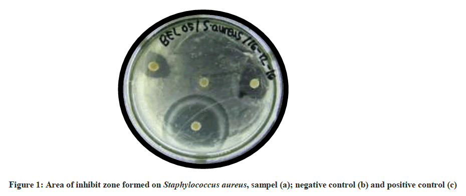
Figure 1: Area of inhibit zone formed on Staphylococcus aureus, sampel (a); negative control (b) and positive control (c)
| No. | Locations | Species of mangrove plants | |||
|---|---|---|---|---|---|
| Rizophora apiculata | Rizophora | Sonneratia alba | Lumnitzera littorea | ||
| sp.1 | |||||
| 1 | Kapo-kapo island | 9 | 4 | 6 | 5 |
| 2 | Setan island | 5 | 7 | - | - |
| Number of isolates | 14 | 11 | 6 | 5 | |
Note: (-): not found this mangrove species in Setan island
Table 1: Endophytic isolates from four species of mangrove plants that produce antibacterial
Thirty-six antibacterial endophytic bacteria were from R. apiculata of fourteen isolates; eleven isolates from Rhizopora sp.1; six isolates from S. alba and five isolates from L. littorea. The presence of different endophytic bacterial isolates in the number of each mangrove species species and based on mangrove living sites located on the two islands, indicating that the presence of endophytic bacterial isolates is influenced by environmental factors as well as from mangrove species itself. Three hundred and seventeen different isolates can be isolated from endophytic bacteria of mangrove plants in Saudi Arabia [8]. Isolation of endophytic bacteria from five mangrove plants (Rhizopora apiculata, Avicennia marina, Exoecaria agallocha, Ceriops decandra and Aegiceras corniculatum) in Pichavaram, Tamil Nadu, India, obtained 104 bacterial colonies with 36 different isolates. Isolated bacteria from Rhizopora apiculata were 26 isolates consisting of 4 isolates and 22 isolates not pigment [3].
Table 2 shows that all endophytic bacteria can produce antibacterial, this is indicated by the inhibition zone (clear zone) with the observation done after 48 hours incubation at room temperature 300°C, formed inhibition zones around the test bacteria, this indicates the ability of endophytic bacterial isolates to produce secondary metabolites in the form of antibacterial compounds that can inhibit bacterial growth. Characterization of metabolites produced by endophytic bacteria, generally done after isolation of bacteria and growing it in vitro [9].
| Location | Species Mangrove / Endophytic Isolate Codes | Diameter of inhibitory zone mm) | |
|---|---|---|---|
| Staphylococcus aureus | Escherichia coli | ||
| Kapo-kapo island | Rhizopora apiculate | ||
| BER-01 | 10 | - | |
| BER-02 | 12 | 14 | |
| BER-03 | 15 | 15 | |
| BER-04 | 7 | - | |
| BER-05 | 14 | 8 | |
| BER-07 | 16 | 8 | |
| BER-09 | - | 10 | |
| BER-11 | 10 | 10 | |
| BER-14 | 20 | 20 | |
| Rhizopora sp.1 : | |||
| BER1-02 | 10 | 10 | |
| BER1-06 | 8 | - | |
| BER1-07 | 15 | - | |
| BER1-09 | - | 10 | |
| Sonneratia alba : | |||
| BES-01 | 12 | 10 | |
| BES-02 | 16 | 18 | |
| BES-05 | 21 | - | |
| BES-07 | - | 8 | |
| BES-08 | - | 24 | |
| BES-09 | 9 | 9 | |
| Lumnitzera littorea | |||
| BEL-01 | 12 | 10 | |
| BEL-04 | 10 | 10 | |
| BEL-05 | 20 | 22 | |
| BEL-07 | 8 | - | |
| BEL-08 | 10 | - | |
| Setan island | Rizophora apiculata | ||
| BER-17 | 15 | 10 | |
| BER-19 | 10 | 10 | |
| BER-21 | 8 | - | |
| BER-23 | 8 | - | |
| BER-24 | - | 12 | |
| Rhizopora sp.1 : | |||
| BER1-10 | - | 10 | |
| BER1-11 | 12 | 12 | |
| BER1-12 | 8 | - | |
| BER1-13 | 11 | 11 | |
| BER1-15 | 14 | 8 | |
| BER1-18 | 7 | 7 | |
| BER1-19 | 8 | 8 | |
Table 2: Diameter of the inhibitory zone of antibacterial various endophytic bacterial isolates of mangrove plants.
Table 2 shows that thirty-six antibacterial-producing isolates with varying antibacterial activity indicated a small diameter of inhibit zone between 7 mm to 22 mm. Thirty isolates of endophytic bacteria have antibacterial activity against S. aureus test bacteria, which are antibacterial of BES-05 isolate with the largest diameter (21 mm), whereas twenty-four isolates have antibacterial activity against E. coli, where isolate BES- 08 is the largest diameter (24 mm). Meanwhile, 20 isolates have antibacterial activity against both test bacteria, where the isolate BEL-05 has the largest diameter (20 mm and 22 mm). The ability of each bacterium is different, in gram-positive single-cell wall structure and low lipid content whereas Gram negative has a triple-cell wall consisting of lipoproteins, phospholipids and lipopolysaccharides and high lipid content [10]. Table 2 also shows that isolate BES-05, including the antibacterial potential isolates against the S. aureus gram-positive bacteria. Potentially these isolates, because the antibacterial they produce are very strong. While isolate BES-08, has a very strong antibacterial category against E. coli gram-negative bacteria. While isolates BER-20 and BEL-05 is a very strong category of antibacterial against both test bacteria and is broad spectrum. The interpretation that is often used in the calculation of the inhibitory namely as follows: the obstacle area with a diameter of 20 mm or more signifies has a very strong potential resistance area with a diameter of 10-20 mm has a strong antibacterial potential obstacle area with a diameter of 5-10 mm has a potent antibacterial potential of the inhibition area with a diameter of 5 mm or less of its weaker antibacterial potential [11]. Based on its activity, antibiotics is divided into two major groups, namely broad-spectrum antibiotics, which are agents that can inhibit growth and kill Gram positive bacteria and Gram negative bacteria, Narrow spectrum is antibiotics target particular types of bacterial such as gram-positive bacteria or gram-negative bacteria [12].
Characterization of endophytic bacteria producing antibacterial
Macroscopic observations of four isolates of endophytic bacteria of mangrove plants were (Table 3): isolate BES-05 and BEL-05 colonies were circular, while isolate BES-08 and BER-20 were irregular colonies. Yellow colored colonies belong to BES-05 and BE-20 isolates, while BES- 08 isolates and BEL-20 colonies are white. The margin of the colony is entire by the isolate BER-05 and BEL-05, while the lobate type is characteristic of the BES-08 and BER-20 isolates. The smooth surface of the colony with elevation flats is characteristic of the isolates BES-05 and BER-20, while isolate BES-08 and BEL-05 with rough and convex characteristics. Microscopic observation showed that all isolates were basil, two isolates were gram-positive (BES-08 and BEL-05) and BES-05 and BER-20 were classified as gram-negative bacteria. Based on macroscopic and microscopic characteristics, the four isolates have different characteristics with each other, indicating that the four isolates are different species of endophytic bacteria (Figure 2).
| Isolate | Macroscopic | Microscopic | |||||
|---|---|---|---|---|---|---|---|
| Colony | Gram | Cell | |||||
| Color | Shape | Margin | Surface | Elevation | stain | shape | |
| BES-05 | Yellow | Circular | Entire | Smooth | Flat | Negative | Basil |
| BES-08 | White | Irregular | Lobate | Rough | Convex | Positive | Basil |
| BER-20 | Yellow | Irregular | Lobate | Smooth | Flat | Negative | Basil |
| BEL-05 | White | Circular | Entire | Rough | Convex | Positive | Basil |
Table 3: Macroscopic and Microscopic of Antibacterial Producing Endophytic Bacterial
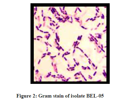
Figure 2: Gram stain of isolate BEL-05
Isolate BES-08 or genus Bacillus sp.1. have differences with isolate BEL-05 genus Bacillus sp.2., seen on MR test and mannitol. BEL-05 isolate showed positive result with marked with medium turning to yellow, while BES-08 isolate was negative. Test with MR medium, showed positive result on BES-08 isolate marked red colored medium, whereas in BEL-05 isolate negative, medium was not red colored. Methyl red test conducted to determine the ability of microorganisms capable of producing organic acid from glucose metabolism.
Methyl red is used to determine the presence of mixed acid fermentation. If there is fermentation of acid mixed broth cultures will remain red. If there is no mixed acid fermentation then the culture broth turns yellow after the addition of methyl red reagent. In methyl red test used methyl red indicator which at the end of observation will show the change of pH to acid. The color change to red means showing the acidic and the yellow discoloration indicates the base color [10].
Physiological characterization and biochemical test (Table 4)
| No. | Biochemical test | Isolate BES-05 | Isolate BES-08 | Isolate BER-20 | Isolate BEL-05 |
|---|---|---|---|---|---|
| 1 | Trypic Soy Agar (TSA) | + | + | + | + |
| 2 | Aerob/Anaerob | + | + | + | + |
| 3 | Gas | - | - | - | - |
| 4 | TSIA | K/K | K/K | K/K | K/K |
| 5 | H2S | - | - | - | - |
| 6 | Catalase | + | + | + | + |
| 7 | Oxidase | - | - | - | - |
| 8 | Motility | + | + | - | + |
| 10 | Indole | - | - | - | - |
| 11 | Urea | - | - | +/- | - |
| 12 | Citrate | + | - | + | - |
| 13 | Lactose | + | - | - | - |
| 14 | Glucose | - | - | + | - |
| 15 | Sucrose | - | - | - | - |
| 16 | Mannitol | +/- | - | +/- | + |
| 17 | MR | +/- | + | - | - |
| 18 | VP | + | +/- | - | +/- |
| 19 | OF | - | - | + | - |
| 20 | KCN | +/- | + | ||
| 21 | Arginine | - | - | ||
| 22 | Lysin | + | - | ||
| 23 | Ornithine | - | - | ||
| 24 | Phenylalanine | - | +/- | ||
| 25 | Aesculin | + | - | ||
| 26 | Arabinose | - | - | - | - |
| 27 | Raffinose | - | - | ||
| 28 | Sorbitol | - | + | ||
| 29 | Threlase | - | - | ||
| 30 | Xylose | - | - | ||
| 31 | Nitrate | ||||
| 32 | Gelatin | + | + | + | + |
| 33 | Dulcitol | - | - | ||
| Species | Enterobacter sp. | Bacillus sp.1 | Klebsiella sp. | Bacillus sp 2. |
Notes: The empty marks on the table show no tests of KCN, arginine, lysine, ornithin, phenylalanine, aesculin, raffinose, sorbitol, threlase, and dulcitol
Table 4: Physiological characterization and biochemical tests of endophytic bacterial mangroves
Conclusion
Five antibacterial bacterial isolates were obtained on mangrove leaf, three isolates had inhibition zone against E. coli bacteria, four isolates had inhibition zone against S. aureus bacteria and two bacterial isolates against both test bacteria. Physiological characterization and biochemical test were obtained for four suspected species, Enterobacter sp., Bacillus sp.1, Klebsiella sp. and Bacillus sp.2.
References
- M.E. Amrani, A. Debbab, A.H. Aly, V. Wray, S. Dobretsov, Tetrahed. Lett., 2012, 53, 6721-6724.
- A. Manilal, T. Tsalla, Z. Zerdo, G. Ameya, B. Merdekios, Asian Pacific J. of Tropical Disease, 2016, 6, (2), 136-140.
- S. Gayathri, D. Saravanan, M. Radhakrishnan, R. Balagurunathan, K. Kathiresan, Indian J. Biotech., 2010, 9, 397-402.
- P. Radell, V. Gordon, Lesson From Nature: Can Ecology Provide New Leads In The Search For Novel Bioactive Chemicals From Rain Forest. Chambridge UK, 2000.
- S.A. Zam, Syamsuardi, A. Agustien, M. Jannah, Y. Aldi, Der Pharm. Lett., 2016, 8(11), 83-89.
- C.A.C. Araujo, L.L Leon, Biological Activities of Curcuma longa L”. Mem Ins Oswaldo Cruz, Rio de Janeiro, 2001.
- A. Djamaan, A. Agustien, D. Yuni, J. Bahan Alam Indonesia., 2012, 8, 1.
- F. Bibi, I. Ullah, S. Akhtar, M. Yasir, E.A. Kensarah, African J. Microbiol. Res., 2017, 11 (19), 729-739.
- G. Brader, S. Compant, B. Mitter, F. Trognitz, A. Sessitch, Curr. Opinion in Biotechnol., 2014, 27, 30-37.
- J.G. Cappuccino, N. Sherman. Microbiology a Laboratory Manual. 5th Edi., Pearson Education, Inc, Publishing as Benjamin Cummings. San Fransisco, 2005.
- W.W. Davis, T.R. Stout, J. Microbiol., 1971, 22(4), 659-665.
- T.G. Slama, A. Amin, S.A. Brunton, AM. J. Med., 2005, 118 (7A), 15-65.


