Research Article - Der Pharma Chemica ( 2017) Volume 9, Issue 1
Electrochemical and Spectroscopic Evaluation on Interaction of Anticancer Drug Bicalutamide with DNA
Umar J Pandit1, Gowhar A Naikoo2, Gulzar A Khan3, Sneha Wankar1, Imran Khan1, Raj KK1 and Limaye SN1*2Department of Mathematics and Sciences, College of Arts and Applied Sciences, Dofar University, Salalah, Oman
3Department of Chemistry, Heterocyclic Synthesis and Electroanalytical Laboratory, Dr. Harisingh Gour (Central) University, Sagar, Madhya Pradesh 470003, India
Limaye SN, Department of Chemistry, Rare Earth and Electroanalytical Research Laboratory, Dr. Harisingh Gour (Central) University, Sagar, Madhya Pradesh 470003, India, Email: admissiondhsgsu@gmail.com
Abstract
Differential Pulse (DP) voltammetry, spectrophotometry, fluorescence emission spectroscopy and docking simulations were employed to investigate the interaction of an anticancer drug Bicalutamide (BIC) with CT-DNA (Calf thymus-DNA). At modified single walled carbon nanotube carbon paste (SWCNT-CP) electrode the anticancer drug BIC produced a well-defined cathodic reduction peak which decreased on addition of CT-DNA. On increasing concentrations of CT-DNA no shift of peak potential of BIC was observed, characteristic of Groove binding mode of interaction. A binding constant (K) of 2.13 × 10⁶ M-1 was determined by DP voltammetric method. In addition, the interaction of BIC with CT-DNA was further examined by spectrophotometric and fluorescent emission techniques. Interestingly, the binding constants (K) obtained through spectrophotometric method and voltammetric technique was in close agreement. However, the groove binding interaction of drug and DNA was also revealed by spectroscopic studies which were supported by theoretical docking studies.
Keywords
Bicalutamide, DNA, Groove binding, Binding constant, Spectrophotometry
Introduction
DNA is an important bio-macromolecule which plays main part in life processes viz storing, copying and transmitting genetic information through genes. From pharmacological point of view, DNA is target for many clinically practical drugs and drugs under advanced clinical trials. The binding nature of these pharmaceuticals with DNA is important in understanding the mechanism of these drugs and also helpful in designing new drugs [1-9].
Various analytical techniques like electrochemical [10-14], spectrophotometry [15], fluorescence [16], NMR [17], FT-IR [18] etc. have been used to study drug-DNA interactions and mechanisms. To understand the drug-DNA interaction mechanism, it is important to introduce techniques which are relatively simple, rapid and more importantly economical. DNA being electrochemically active with many other drug molecules exhibit redox activity. These electrochemical methods especially cyclic voltammetry, differential pulse voltammetry and stripping voltammetry have been widely employed to understand the mechanism of DNA-drug interactions due to their high sensitivity and economical nature.
Many binding mechanism are operative in drug-DNA interaction, the important of which are intercalative binding, covalent binding, non-covalent binding and external binding [3,19]. Among these, the intercalative binding which can occur as classical intercalation, threading intercalation or through groove binding results in insertion of aromatic ligand into adjacent base pairs on the DNA, which results in distortion of DNA structure and thus thwarting its biological function [2,3]. The study of the drug-DNA mechanisms have
considerably increased the knowledge for better understanding the pharmacokinetics of many anti-cancer drugs with the advent of developing more powerful DNA targeted pharmaceuticals.
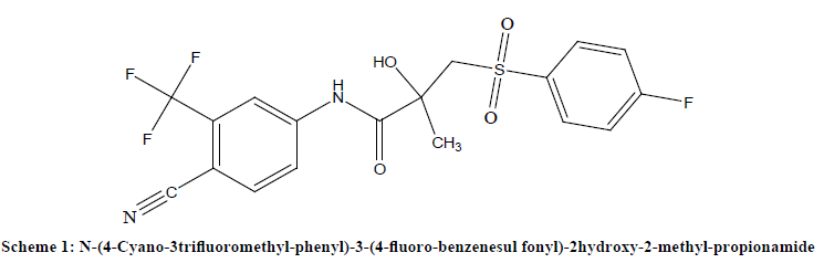
The present work reveals the investigation on the interaction of an anticancer drug Bicalutamide (BIC) [N-(4-cyano-3- trifluoromethyl-phenyl)-3-(4-fluoro-phenylsulfonyl)-2-hydroxy-2-methyl-propionamide (Scheme 1)] with calf-thymus DNA (CTDNA) by voltammetric technique along with the support of fluorescence, spectrophotometric techniques followed by theoretical studies.
Chemical and materials
CT-DNA, Bicalutamide pure, graphite powder and SWCNT’s (Sigma-Aldrich) were used without further purification. All other chemicals and reagents used were of analytical grade and purchased from Merck India. Stock standard solution of BIC was prepared by dissolving 1 mg BIC in 10 ml ethanol solution. 0.1 M phosphate buffer was prepared in ultrapure water. CT-DNA solution was prepared in 0.1 M phosphate buffer of pH 7.0 ± 0.2 by dissolving 1 mg CT-DNA in 100 ml buffer solution. The concentration of stock solution of CT-DNA was determined by spectrophotometry using molar absorption coefficient of 6600 cm-1M-1 [20]. Working solutions of drug and CT-DNA were prepared by diluting standard solutions with appropriate volumes of solvent or buffer solutions.
Preparation of modified carbon paste electrode
Bare and modified Carbon Paste Electrodes (CPE) was prepared according to literature reports [21,22] by hand-mixing Graphite powder and mineral oil (paraffin oil) in 70:30 ratios for bare CPE. Modified SWCNT-CPE was prepared in the same way by mixing SWCNT’s: Graphite: Paraffin oil in the ratio 10:60:30. The resulting paste was filled in polyethylene syringe of 2 mm internal diameter with a pre-inserted copper wire to establish an external electric contact. Fresh electrode surface for each measurement was generated by mechanically pressing the paste from top and smoothed using weighing paper. Finally the electrodes were carefully washed with distilled water.
Instrumentation
Voltammetric measurements were performed with a computer-controlled Electrochemical Ion Analyzer model Ω 797 VA Computrace (Swiss made), assembled with a three electrode cell employing a bare and hand-made SWCNT-CP working electrode, an Ag/AgCl (saturated KCl) reference electrode and a platinum wire as counter electrode. Spectrophotometric experiments were carried out on Systronics 2201 double beam UV-Vis spectrophotometer. Fluorescence spectra were recorded on Shimadzu RF 5301 spectrofluorometer equipped with a Xe lamp as the excitation source. Excitation and emission slits were 5 nm each with the instrument operating in high sensitivity and super scan speed. All pH measurements were performed with Systronic digital μpH meter model-361. All experiments were performed at room temperature. Pure nitrogen gas was purged through test solutions for 5 minutes for oxygen free atmosphere.
Conclusion
The work describes interaction of anticancer drug bicalutamide with calf thymus-DNA. Electrochemical and spectroscopic methods were used to study the drug-DNA interaction. The results were used to elucidate the binding constant (K) and Stern-Volmer quenching constant (KSV). The slight higher value of binding constant (K) obtained through voltammetric technique over that obtained in spectrophotometric technique may be a consequence of higher sensitivity of electrochemical techniques. The Stern-Volmer quenching constant obtained in Fluorescence emission spectroscopy, supports that BIC is an efficient DNA intercalating agent. Electrochemical technique suggested intercalative mode of interaction between BIC and DNA, while spectroscopic techniques proved that groove-binding type of interaction is mainly operating between the drug and CT-DNA. The groove-binding mode of interaction was also supported by docking simulation studies.
Acknowledgments
Authors are highly thankful to UGC-SAP for electrochemical instrumentation facilities. Authors also acknowledge Department of Pharmaceutical sciences and Department of Chemistry, Dr. Harisingh Gour (Central) University Sagar (M.P.) India for providing necessary laboratory facility.
References
[1] X. Lu, Y. Chen, J. Chen, Y. Zhang, L. Zhang, M. Li, Int. J. Electrochem. Sci., 2006, 1, 130.
[2] Q. Wang, X. Wang, Z. Yu, X. Yuan, K. Jiao, Int. J. Electrochem. Sci., 2011, 6, 5470.
[3] M. Sirajuddin, S. Ali, A. Badshah, J. Photochem. Photobio. B: Biol., 2013, 124, 1.
[4] X. Tian, Y. Song, H. Dong, B. Ye, Bioelectrochemical., 2008, 73, 18.
[5] L. Wang, L. Lin, Y. Baoxian, J. Pharm. Biomed. Anal., 2006, 42, 625.
[6] S.S. Kalanur, U. Katrahalli, J. Seetharamappa, J. Electroanal. Chem., 2009, 636, 93.
[7] C. Xia, L.S. Guo, J.H. Jiang, T.F. Kang, B. Xiong, Y. Ru-Qin, Anal. Chim. Acta., 1998, 373, 29.
[8] S.C.B. Oliveira, A.M. Chiorcea-Paquim, S.M. Ribeiro, A.T.P. Melo M. Vivanc, A.M. OliveiraBrett, Bioelectrochemical., 2009, 76, 201.
[9] S. Raufa, J.J. Gooding, K. Akhtar, M.A. Ghauria, M. Rahman, M.A. Anwar, A.M. Khalid, J. Pharm. Biomed. Anal., 2005, 37, 205.
[10] B.X. Ye, L.J. Yuan, C. Chen, J.C. Tao, Electroanalysis, 2005, 17, 1523.
[11] G. Congur, A. Erdem, F. Mese, Bioelectrochemistry, 2015, 102, 21.
[12] A.D.R. Pontinha, S. Sparapani, S. Neidle, A.M. Oliveira-Brett, Bioelectrochemistry, 2013, 89, 50.
[13] X. Tian, F. Li, L. Zhu, B. Ye, J. Electroanal. Chem., 2008, 621, 1.
[14] F.P. Zhang, Q. Cao, J.J. Cheng, C.H. Zhang, N. An, S.P. Bi, Anal. Sci., 2009, 25, 1019.
[15] C.V. Kumar, J.K. Barton, N.J.J. Turro, J. Am. Chem. Soc., 1985, 107, 5518.
[16] C.V. Kumar, E.H. Asuncion, J. Am. Chem. Soc., 1993, 115, 8547.
[17] L. Scaglioni, S. Mazzini, R. Mondelli, S. Dallavalle, S. Gattinoni, S. Tinelli, G.L. Beretta, F. Zunino, E. Ragg, Bioorg. Med. Chem., 2009, 17, 484.
[18] S.T. Saito, G. Silva, J. Photochem. Photobio., B: Biol., 2012, 111, 59.
[19] X.H. Zhang, L.Y. Wang, Z.X. Nan, S.H. Tan, Z.X. Zhang, Dyes and Pigments., 2008, 79, 205.
[20] R.A. Dar, P.K. Brahman, S. Tiwari, K.S. Pitre, J. Appl. Electrochem., 2011, 41, 1311.
[21] B.H.M. Hussein, H.A. Azab, W. Fathalla, S.A.M. Ali, J. Luminescence, 2013, 134, 441.
[22] J. Wang, Analytical Electrochemistry, Willey, NY, USA, 2001, 2nd Ed., 115.
[23] N.P. Shetti, S.J. Malode, S.T. Nandibewoor, Bioelectrochemistry, 2012, 88, 76.
[24] U.J. Pandit, I. Khan, S. Wankar, K.K. Raj, S. N. Limaye, Anal. Methods., 2015, 7, 10192.
[25] M.T. Carter, A.J. Bard, J. Am. Chem. Soc., 1987, 109, 7528.
[26] N. Li, Y. Ma, C. Yang, L. Guo, X. Yang, Biophys. Chem., 2005, 116, 199.
[27] H. Nawaz, S. Rauf, K. Akhtar, A.M. Khalid, Anal. Biochem., 2006, 354, 28.
[28] F. Jalali, P.S. Dorraji, J. Pharm. Biomed. Anal., 2012, 70, 598.
[29] J. Liu, T. Zhang, T. Lu, L. Qu, H. Zhou, Q. Zhang, L. Ji, J. Inorg. Biochem., 2002, 91, 269.
[30] J. Jaumot, R. Gargallo, Curr. Pharmaceut. Des., 2012, 18, 1900.
[31] Y.J. Guo, J.B. Chao, J.H. Pan, Spectrochim. Acta. Part A, 2007, 68, 231.
[32] H.A. Benesi, J.H. Hildebrand, J. Am. Chem. Soc., 1949, 71, 2703.
[33] J.R. Lakowicz, Springer, NY, USA, 2006, 3rd ed., 241.
[34] R.D. Taylor, P.J. Jewsbury, J. W. Essex, Journal Computer Aided Drug Design., 2002, 16, 151.
[35] H.R. Drew, R.M. Wing, T. Takano, C. Broka, S. Tanaka, K. Itakura, R.E. Dickerson, Proc. Natl. Acad. Sci. USA., 1981, 78, 2179.
[36] O. Trott, A.J. Olson, J. Comput. Chem., 2010, 31, 455.
[37] Accelrys Software Inc., Release 4.0, San Diego: Accelrys Software Inc., 2013.


