Review Article - Der Pharma Chemica ( 2022) Volume 14, Issue 8
Phytochemical, Pharmacological and Toxical Proprieties of Euphorbia Helioscopia: Review
Anjoud Harmouzi1,2*, Yassine El Ammari1, Amina CHLOUCHI1, Rabha AFROUKH1 and Ahmed Boughdad22Department of Plant Protection and Environment. National School of Agriculture - Meknes. B.P. S/40 50000; Meknes, Morocco
Anjoud Harmouzi, Agrophysiology, Biotechnology, Environment and Quality laboratory, Sciences Faculty, Ibn Tofail University, BP 133, 14000 Kenitra, Morocco, Email: nojoud.harmouzi@gmail.com
Received: 22-Jul-2022, Manuscript No. dpc-22-70003; Editor assigned: 25-Jul-2022, Pre QC No. dpc-22-70003; Reviewed: 08-Aug-2022, QC No. dpc-22-70003; Revised: 11-Aug-2022, Manuscript No. dpc-22-70003; Published: 26-Aug-2022, DOI: 10.4172/0975-413X.14.8.1-35
Abstract
Euphorbia helioscopia L. (Euphorbiaceae), also known as “Sun spurge” is one of used herbs in most parts of the world since thousands of years. This work aims to provide updated comprehensive and categorized information on chemical composition, pharmacological and toxical properties of this plant. To achieve this purpose, several scientific databases were used, including Wiley, PubMed, Web of Science, Springer, Science Direct, Elsevier, and Google Scholar and related books published up to December 2019 were also used as references. After description of its history, botanical characteristics and distribution, this review highlights the different types of secondary metabolites, such as diterpenoids, tannins, glucosides and steroids, which have been isolated from E. helioscopia L. in several researches over the last four decades. Related Twenty five pharmacological activities (among them: antibacterial, antiviral, antioxidant, antifungal, phytotoxic, anthelmintic, antitumor, anti-allergic, anti-asthmatic, cytotoxic, anticancer, allelopathic, anti-nociceptive, anti-inflammatory, anti-pyretic, vasodepressor, antileishmanial, lipid-lowering activity, and triglyceride-lowering activity) are presented and discussed. E. helioscopia L. toxicity related studies against Humans, snails and some herbs are summarised and presented with correspondent references. This study can be used by researches interested by this plant since it resumes all relevant published papers in link with E. helioscopia L.
Keywords
Euphorbia Helioscopia; Chemical composition; Pharmacological properties; Toxicity
INTRODUCTION
Euphorbiaceae is the largest family of Anthophyta with 300 genera and 5,000 species [1]. Euphorbia, is one of the largest angiosperms’ genera, with about 2,000 species, and has long been known for its wide variety of growth forms, including many xerophytic species. Despite its great vegetative diversity, the genus is morphological united by the possession of a cyathium which is a very small inflorescence that looks like a single flower [1-3].
Euphorbia helioscopia L., also called Sun spurge, is an annual plant with a tall between 10 and 50 cm high, with a rich latex stem, oval alternate leaves and small yellow-green flowers. The plant is native to temperate regions of Eurasia but has been adapted to subtropical conditions. It behaves as a winter annual in Japan that is blooming from April to May. In Morocco, this plant blooms from December to April in plains and up to May in mountainous areas [4]. Most E. helioscopia L. compounds are biologically active; for example: Antibacterial [1, 5-8], antitumor/anticancer [9-12] antioxidant [1, 13-15], Antifungal [16] plus other types of activities. Plants in Euphorbiaceae family are also promising sources of high quality tanning agents for leather industry [17]. E. helioscopia L. has been considered as a medicinal plant and is used in traditional medicine in various countries of the world [3, 18-20]. For Maghrebian traditional medicine, E. helioscopia L. is locally called "Bou Labeena" (in reference to the plant’s milky latex). However, Euphorbia stems are an irritant drug that cause severe toxicity like phytotoxic effects [21], Molluscicidal activity [22-24] and skin irritant [25].
The objective of this review is to present an evaluation of art state of E. helioscopia L., in particular chemical compounds identified and characterized in its different parts, in one hand; in the other hand, to examine pharmacological and toxic properties of this plant, based on a comprehensive and in-depth analysis of relevant literature.
MATERIALS AND METHODS
In this work we collected literatures published documents up to December 2019 on phytochemistry, botany, pharmacology and toxicity of E. helioscopia L. All available information on this plant was retrieved by Internet databases (Springer, PubMed, Web of Science, Wiley, Science Direct, Elsevier, and Google Scholar) and a number of books and PhD dissertations were also used. Used key words in the search were: “Euphorbia helioscopia L.”, “Traditional uses of Euphorbia helioscopia L.”, “Chemical composition of Euphorbia helioscopia L.”, “pharmalogical activity”, “toxicity”. Scientific name of the plant has been identified and validated by the Scientific Institute of Rabat (Morocco) and through www.theplantlist.org.
Traditional uses of Euphorbia L. (Euphorbiaceae)
Historically, natural extract from plants have showed a significant role in humans live, whether for food or for drugs [26]. Euphorbia L. (Euphorbiaceae) has attracted human interest around the world since the earliest and even prehistoric times [27-29]. Euphorbia species are easily identified especially by their milky latex and unique inflorescences (cyathia) [30]. They are known for their use as ornamental and domestic plants [31], and their latex has contributed to the economic interest for certain species such as Euphorbia antisyphilitica Zucc [31] and Euphorbia Intisy Drake (intisy rubber) [31]. According to Hippocrates, Galen and Dioscorides [32, 33], Sumerians and Akkadians knew the toxic properties of E. helioscopia L. [34], the medicinal properties of "euphorbium" (gum from E. resinifera Berg [32] and the presence of active natural products in the latex [35,36]. In 1752 Carl Linnaeus presented Euphorbia genus with various medicinal uses, including topical treatments and purging of the gastrointestinal tract [37-39]. E. helioscopia L. takes her name from a Greek origin, deriving from ‘‘helioscopion’’ used first by Dioscorides to describe this species and meaning ‘‘sun gazer’’. In fact, according to the Roman naturalist Pliny, the plant moves its flowers round to follow the sunlight [40, 41]. Furthermore, as reported for other species belonging to the Euphorbiae genus, the plant was used by Greeks and Romans to take away all kinds of warts, knobs, and the hard calluses of fistulas, hot swelling and carbonucles. Nowadays, the plant is still used in China for a variety of ailments: as a cure for boils, to get rid of worms, and to bring down high temperatures [41].
Botanical characteristics
E. helioscopia L. (Figure 1) is a fleshy annual plant, glossy green and glabrous, up to 50 cm tall but more commonly between 10 to 30 cm. All parts of the plant emit a milky sap, once cut. E. helioscopia L.’s stems are clearly tinged with red and radiate outward from a central point. All leaves (1 to 2 mm long) are usually attached directly to the stem and do not appear on petioles. Not very visible, light green to pale yellow, inflorescences (cyathias) occur at stems’ ends and have 4 lobes. Each lobe has a round yellowish gland [42, 43].

Figure 1: Euphorbia helioscopia L. (By Anjoud HARMOUZI)
E. helioscopia L. is classified in reign: Plantae, under reign: Tracheobionta, Super division: Spermatophyta, Division: Magnoliophyta, Class: Magnoliopsida, Subclass: Rosidae, Order: Malpighiales, Family Euphorbiaceae, Genus: Euphorbia and Species: Euphorbia helioscopia [43].
Distribution
Euphorbia helioscopia is native to North Africa, Europe, and some parts of Asia. It was introduced in North and South America as shown in the cart below (Figure 2). African native countries are Algeria, Egypt, Libya, Morocco and Tunisia, European ones are: Albania, Belgium, Bulgaria, Cyprus, Czech, Finland, France, Germany, Great Britain, Greece, Hungary, Ireland, Italy, Netherlands, Norway, Poland, Portugal, Romania. Asian ones are: Afghanistan, Bangladesh, Belarus, China, India, Iran, Iraq, Japan, Kazakhstan, Kirgizstan, South Korea, Lebanon, Syria, Mongolia, Pakistan, Palestine, Saudi Arabia, Tadzhikistan, Taiwan, Turkey, Turkmenistan and Vietnam. While introduced countries are: Canada, United States of America and Argentina [44-52].

Figure 2: Distribution of Euphorbia helioscopia L. (https://www.ipni.org/n/346754-1)
Chemical composition
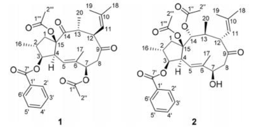
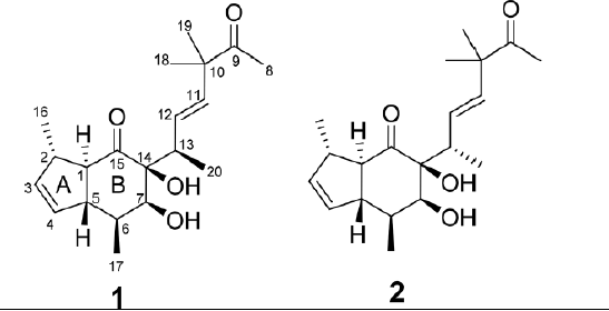
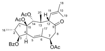
Euphorbia is the largest genus of Euphorbiaceae family, which is well known for the chemical diversity of terpenoids [35]. Diterpenoids are particularly rich and diversified in Euphorbia species, with more than 650 diterpenoids that have been isolated and identified, including jatrophanes, lathyranes, ingenanes, tiglianes, abietanes, kauranes, pimaranes, daphnanes, casbbanes and myrsinanes [35- 36, 53]. A large number of secondary metabolites have been reported from E. helioscopia L. plant including diterpenoid, triterpenoid, tannins, glucosides and lipids [3,54-59] which offer to E. helioscopia L. a wide array of bioactive functions.
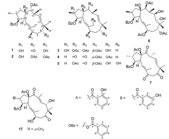
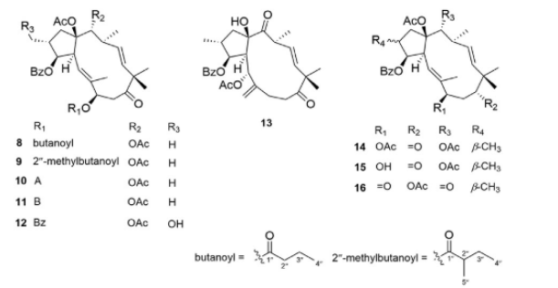
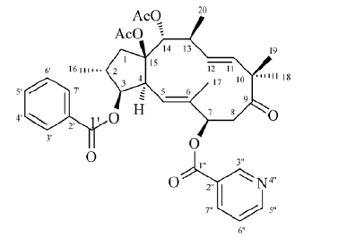
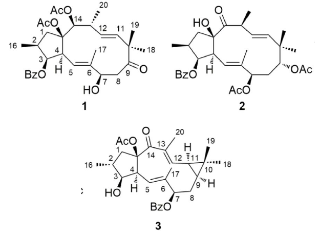
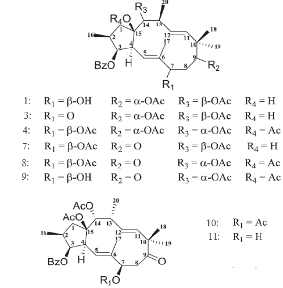
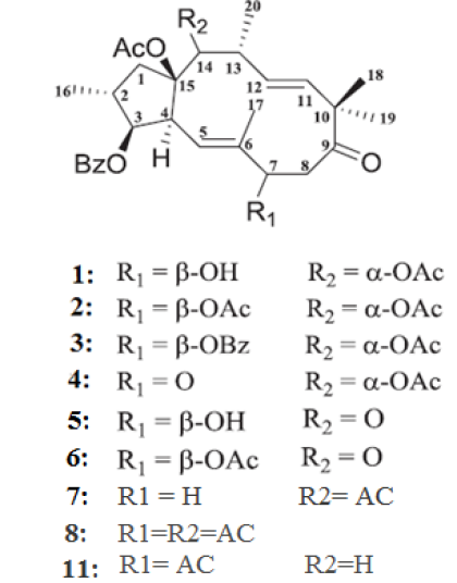
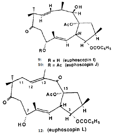
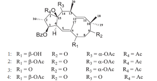
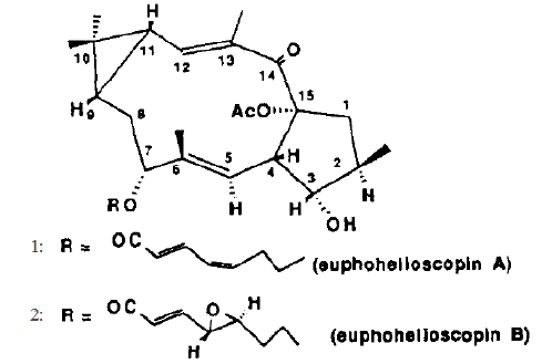
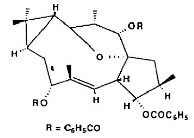
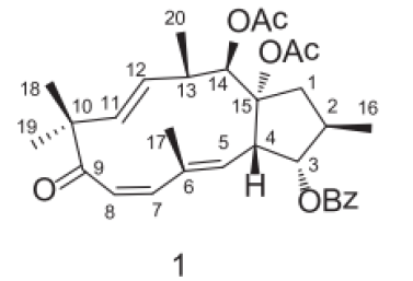
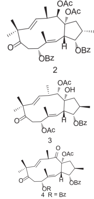
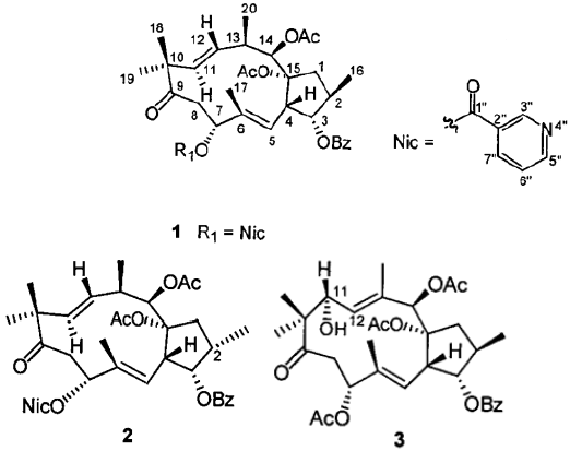
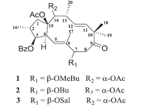
More than 133 diterpenoids have been isolated and structurally characterized from E. helioscopia L. [57, 60], which present the main class components studied in this plant (table 1), e.g Euphohelin A [61], Euphornin A-K (1-11), Euphoscopin A-L, Euphorpin A- F [62], Euphorbiapene A-D [63, 62] and Helioscopianoids A–Q [64].
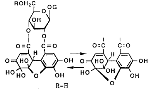
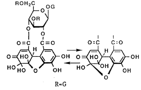
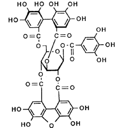
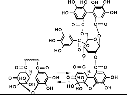
Oil content of Euphorbia helioscopia’s seeds is equal to 30.33 % with a d15: 0.9346, nD: 1.4847, Iodine index, % J2: 176.6–204.4, and saponification value is 191.1mg KOH [65]. According to [64], E. helioscopia L. contains diterpenoid esters of jatrophane, helioscopianoides A–Q and euphornin N [66-67, 115]. Triterpenes like Euphorbatrine A-G [68]. Four hydrolysable tannins named: heliscopinins A & B, and helioscopins A & B were found in all parts of E. helioscopia L. [55-17, 69]. Other studies have revealed the presence of flavonoids like, Heliosin [70,71], glycosides such as quercetin-3-pglucoside, quercetin-3-β-galactoside, quercetin-3-β-galactoside-2"-galla [72] and aryl glycoside, 300-O-galloyl-benzyl-O-α-L-rhamnopyranosyl-(1→6)-β-D-glucopyranoside [57-73,74], Hyperoside and Quercetin [75], steroids and lipids like Glucoclionasterol [54, 59] and other secondary metabolites such as 24-methylene cycloartanol, 24-methylenecycloart-3-one, cycloartanol, and stigmast-4-ene-3-one [3]. Diversity in secondary metabolites in all parts of E. helioscopia L. would explain their various uses in several areas.
| Compound class | Compound name and formula | Chemical structure | References |
|---|---|---|---|
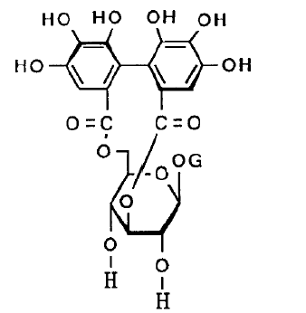
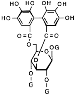
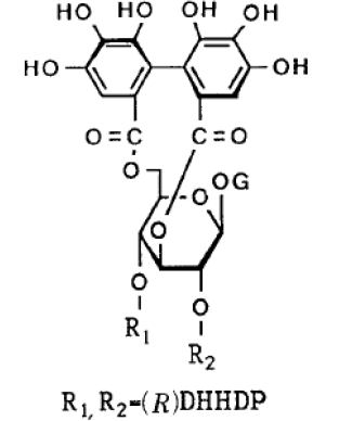
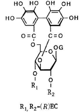
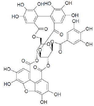
| Tannins | Helioscopin A C47H34O32 |
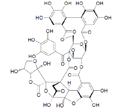 |
[17-55,69] |
| Helioscopin B C47H36O32 |
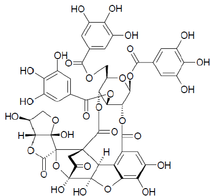 |
[17-55,69] | |
| Helioscopinin A C41H28O27 |
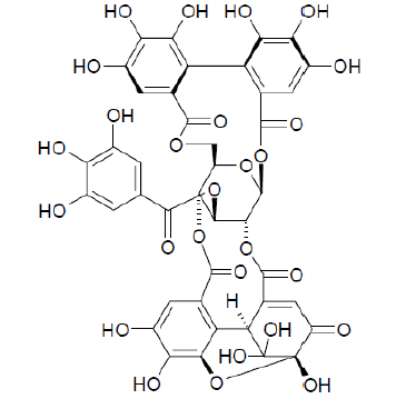 |
[17-55-69, 76] | |
| Helioscopinin B C27H22O18 |
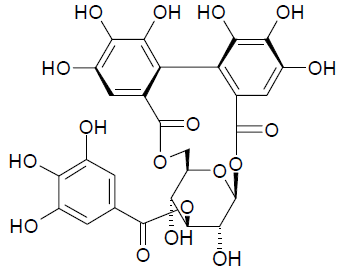 |
[17-55,69] | |
| Corilagin C27H22O18 |
 |
[55] | |
| Punicafolin C41H30O26 |
 |
[55] | |
| Geraniin C41H28O27 |
 |
[55] | |
| Elaeocarpusin C47H34O32 |
 |
[55] | |
| Furosin C27H22O19 |
 |
[55] | |
| Terchebin C41H30O27 |
 |
[55] | |
| Mallotusinin C41H26O25 |
 |
[55] | |
| Carpinusin C41H30O27 |
 |
[55] | |
| Mauotusinin C41H26O25 |
 |
[55] | |
| Diterpenoids | Helioscopinolide A C20H28O3 |
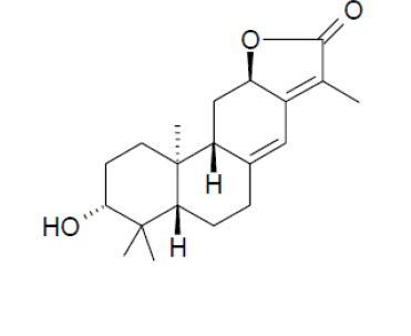 |
[69] |
| Helioscopinolide B C20H28O3 |
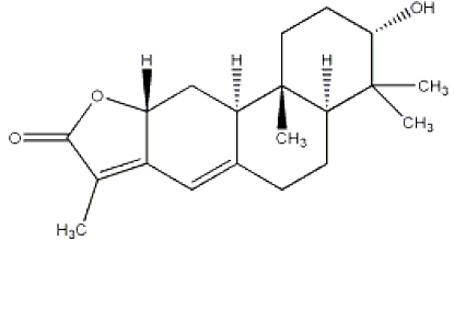 |
[69] | |
| Helioscopinolide C C20H26O4 |
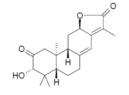 |
[69] | |
| Helioscopianoids A–Q 1: C29H38O7Na 2: C33H44O9Na 3:C33H44O10Na 4: C31H38O8Na 5: C31H40O9Na 6:C33H46O11Na 7: C29H34O7Na 8: C35H46O9Na 9: C36H48O9Na 10: C38H44O10Na 11: C38H44O10Na 12: C38H44O10Na 13:C29H36O7Na 14:C33H42O9Na 15:C31H40O8Na 16:C31H38O8Na 17:C29H36O8Na |
 |
[64] | |
| Euphohelin A C33H44O11 Euphohelin B C31H42O10 Euphohelin C C40H48O12 Euphohelin D C31H38O10 |
 |
[61] | |
| Euphornin C33H44O91 | 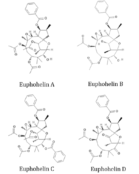 |
[10-54-62-63-66- 67,115] | |
| Euphoheliosnoid E C37H43NO9 | 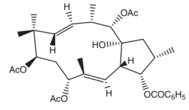 |
[77] | |
| Euphoscopoids A-C
(1-3)
1: C31H40O8 2 : C31H40O8 3 : C29H36O6 |
 |
[53] | |
| Euphornin A-K (1-11) 1 : C31H41O7 2 : C31H42O7 3 : C31H40O8 4 : C35H46O10 5 : C35H46O10 6 : C29H38O7 7 : C31H40O8 8 : C33H42O9 9 : C33H42O9 10 : C31H40O8 11 : C31H40O8 |
 |
[54-62, 78] | |
| Euphoscopin A-L (1-12)
1: C31H40O8 2: C33H42O9 3: C38H44O9 4: C31H38O8 5: C29H36O7 6: C31H38O8 7 :C29H38O7 8: C31H40O8 9: C29H38O7 10: C31H40O8 11: C29H39O7 12: C29H36O7 |
 |
[54,62] | |
| Epieuphoscopin A-B (1-2)
1 : C31H40O8 2: C33H42O9 Epieuphoscopin D (3) 3: C31H38O8 Epieuphoscopin F (4) 4: C31H38O8 |
 |
[54,62] | |
| Euphohelioscopin A-B (1-2) 1: C30H42O6 2 : C30H42O7 |
 |
[54] | |
| Euphohelionon C14H44O7 |
 |
[54] | |
| Euphorbiapene A-D
(1-4) 1: C31H38O7 2 : C38H48O9N 3 : C31H41O8 4 : C36H40O8Na |
 |
[63, 62] | |
| Euphoheliosnoid A-C (1-3)
1: C37H43NO9 2 : C37H43NO9 3 : C33H42O10 |
 |
[79] | |
| Euphorpin A- F (1-6)
1: C36H48O9Na 2: C35H46O9Na 3: C38H44O10Na 4: C29H36O7Na 5-6: C33H42O8Na |
 |
[62] | |
| Monodeacetyleuphornin (C31H42O8Na) |
 |
[78] | |
| Euphornoate A-U (1-21)
1: C34H46O9Na 2: C33H43ClO9Na 3: C34H44O9Na 4: C35H46O9Na 5: C35H48O9Na 6: C37H52O9Na 7: C38H52O9Na 8: C38H46O9Na 9: C38H45ClO9Na 10: C38H45FO9Na 11: C38H45NO11Na 12: C39H45F3O9Na 13: C39H48O9Na 14: C39H48O9Na 15: C39H48O9Na 16: C39H48O10Na 17: C40H50O11Na 18: C39H48O9Na 19: C40H48O9Na 20: C36H44O9SNa 21: C36H44O10Na |
 |
[78] | |
| Heliojatrone A-B (1-2) 1 :C31H38O8 2 : C31H40O8 |
 |
[80] | |
| Euphorhelipane A-B
(1-2) 1: C20H30O4 2: C20H30O4 |
 |
[81] | |
| Heliojatrone C C33H42O9Na |
 |
[82] | |
| Euphcopenoid A-B
(1-2) 1: C20H26O4 2: C20H28O4 |
 |
[83] | |
| Euphoscopoid D-F (1-3) 1: C29H34O7Na 2: C31H40O7Na 3: C36H40O8N |
 |
[82] | |
| Jatrophane-type diterpenoids (1-4) 1: C33H44O10 2: C29H40O7 3: C33H44O10 4: C38H44O10 |
 |
[11] | |
| Euphorhelipanes A-B (1-2) 1: C20H30O4 2: C20H30O4 |
 |
[8] | |
| Heliosterpenoids A-B (1-2) 1: C31H38O7Na 2 : C29H36O6Na |
 |
[84] | |
| Secoheliosphanes A and B (1-2) 1: C31H40O8 2: C31H40O8 |
|
[85] | |
| Secoheliospholane A C31H40O10 |
|
[85] | |
| 2-epi-Euphornin I C31H40O8 |
|
[85] | |
| Euphelionolide A-N (1-14) 1-7: C20H28O5Na 8-9 : C20H26O5Na 10-11 : C20H26O6Na 12-13 : C20H28O5Na 14 : C20H26O5Na |
|
[86] | |
| 6-Epi-18-hydroxy-abbeokutone C20H32O4Na |
|
[86] | |
| Eupheliotriol A-B
(1-2) 1: C20H30O4Na 2: C20H30O5Na |
|
[86] | |
| Triterpenes | Euphorbatrine A-G (1–7)
(19αH)-lupane : (1-2) : 1 : C29H48O2 2 : C30H48O2 3 : C29H46O2 (9βH)-lanostane : (4–6) 4 : C30H50O2 5 : C30H50O2 6 : C30H48O2 7 : C30H46O3 |
|
[68] |
| Flavonoids | Heliosin C27H30O17 |
|
[56, 71] |
| Falavones | Quercetin-3-O-glucoside C21H19O12 |
|
[87] |
| Flavonols | Kaempferol C15H10O6 |
|
[88] |
| Flavonol glycosides | Isomyricitrin C21H20O13 |
|
[75] |
| Glucosides | Hyperoside C21H20O12 |
|
[56, 75] |
| Quercetin C15H10O7 |
|
[56, 75] | |
| Quercetin-3-O-β, galactoside-2" –galate C28H24O16 |
|
[87] | |
| Titimalin (Quercetin-5,3-digalactoside) C27H30O17 |
|
[70-89, 90] | |
| Glycosphingolipides | Cerebrosides 1-7 C41H77NO9 |
|
[59] |
| Galactolipids | Digalactosyldiacylgycerol | |
[59] |
| Monogalactosyldiacylglycerol C43H76O10 |
|
[59] | |
| Lipids | Glucoclionasterol C35H60O6 |
|
[59] |
Details related to extraction methods used can be found in mentioned references. Throughout this review, it is seen that chemical proprieties are deeply investigated by researchers from different countries from 1968 up to nowadays (2019). Some substances are present in all parts of the plant while other are located in some of them (table3). Further studies about the use of these chemical constituents can be envisaged. E. helioscopia L. diterpenoids have been extensively studied over the years. However, for many other classes of compounds, with have nteresting pharmacological affects possibilities, investigations are scarce. Examples of these compounds include saponins especially in E. helioscopia L. roots, and tannins isolated from E. helioscopia L. where there is still much to be explored and published.
Pharmacological properties
In literature, twenty five pharmacological activities (table 2) were reported in relation with E. helioscopia L. References of studies describing these activities are summarized in table 3 below and are chronologically classified as described in the literature.
| Pharmacological activity | References |
|---|---|
| Antibacterial activity | [1-5-7-77-91,93] |
| Antiviral activity | [7,85] |
| Antioxidant activity | [1-13-14,15] |
| Antifungal activity | [1-16,95] |
| Phytotoxic activity | [1] |
| Anthelmintic activity | [8, 96] |
| Antitumor activity | [9,68] |
| Anti-allergic activity | [76] |
| Anti-asthmatic activity | [76] |
| Cytotoxic activity | ([10-11-53,84] |
| Anticancer activity | [12-41-92-91,94] |
| Allelopathic activity | [98,99] |
| Anti-nociceptive activity | [15] |
| Anti-inflammatory activity | [15,82] |
| Anti-pyretic activity | [15] |
| Vasodepressor activity | [3] |
| Free-radical scavenging activity | [59] |
| Antifeedant | [53] |
| Inhibitory activity | [63] |
| Antileishmanial Activity | [100] |
| Catalytic activity | [101] |
| P-glycoprotein (P-gp) inhibitory | [84] |
| Lipid-lowering activity | [62] |
| Multidrug resistance modulators activity | [78] |
| Triglyceride-lowering activity | [8] |
The most interesting active compounds from E. helioscopia L. belongs to diterpenoids [9, 12]. For example, the bioactive jatrophane diterpenoides, heliosterpenoids A & B were found to be potent inhibitors of P-glycoprotein (ABCB1) and exhibited cytotoxicity against MDA-MB-231 cell lines [85]. The same Helioscopianoids A–Q showed neuroprotective activities against serum deprivation-induced PC12 cell damage [80], euphelionolides F and L diterpenoids exhibited significant cytotoxicity against MCF-7 and PANC-1 cell lines [86]. Triterpenoids Euphorbatrine E and F increased Hela−/− cell death via activating apoptosis pathway other than necroptosis [68]. And the most relevant activities are antibacterial [1-5-7-66-91, 93], antioxidant [1-13-14, 15] and anticancer [12-41-92-91, 94]. As previously described, E. helioscopia L. has several properties, among them its antioxidant activity E. helioscopia L. flowers extracts have the highest levels of phenolics and flavonoids, as well as highest antioxidant compared to leaf and stem extracts, regardless of the solvent used [14].Another study reported that methanolic extracts of E. helioscopia L. leaves possess significant antioxidant activity in vivo by increasing biomarkers of oxidative stress in tissue and homogenates serum enzymes [15].
Among bioactive functions of E. helioscopia L., some studies mentioned an antiviral and antibacterial effect [6-7, 8]. In addition, it has been reported that antibacterial activity of dichloromethane and methanol extracts from aerial parts of E. helioscopia L. was performed against Eschericha Coli, Bacillus Subtilis, Shigella Flexenari, Staphylococcus Aureu, Pseudomonas Aeruginosa and Salmonella Typhi [1]. E. helioscopia L. was active against Bacillus Anthracis [5]. As well as extracts from E. helioscopia L. had an inhibition of more than 90% against Botrytis Cinerea, Rhizoctonia, Solani, Fusarium Oxysporum, Cladosporium Cucumerinum and Alternaria Solani Fungi [16].
Isolated compounds (Euphornin L. and Euphoscopin) of E. helioscopia L. exhibited cytotoxicity against HL-60 [10]. Extracts of E. helioscopia L. from Turkey showed a vasodepressor effect which could be due to its vasorelaxant activity [3]. E. helioscopia L. also showed an anticancer effect; indeed, ethyl acetate and chloroform extracts could inhibit the proliferation of five human cancer cell lines over a range of concentrations from 50 to 200 μg / mL [12]. Park's study [76] indicates that polyphenol compound known as helioscopinin-A has shown some inhibitory activity of capillary permeability in responses to passive cutaneous anaphylaxis in rats. The whole plant of E. helioscopia L. has significant anthelmintic activity, which may be a potential alternative to treating helminth infections in ruminants [96]. Studies showed that E. helioscopia L. have innumerable benefits especially from aerial parts, such as antioxidant, antiviral, antitumoral, antibacterial, antifungal, anti-allergic, anti-asthmatic allelopathic, lipid and triglyceride lowering activity and other effects, it is very promising as anthelmintic.
Table 3 below provides a summary of reported pharmacological activities, details of extracts, application type (in vivo or in vitro), test objective, used control; doses tested for drugs and found results. Among the thirty nine studies about pharmacological activities, 40 % of presented tests were conducted in vivo, 10% of them are performed in vivo and in vitro. Where extracts from all parts of the plant were used to determine these pharmacological proprieties. Encouraging findings can be considered as a scientific demonstration of the different historical uses of this plant as drug as specified in the chapter 3 of this review.
All previously cited researchers used the whole plant of E. helioscopia L. for their studies. However, there are no studies that use each organ (roots, stems, leaves or flowers) of the plant separately and compare their effects for example. The same remark is applicable for comparison of different methods of extraction or solvents that give the best yield extracts. Studies show that E. helioscopia L countless health benefits, such as antibacterial, antiviral, antioxidant, antifungal, anthelmintic, antitumor, anti-allergic, anti-asthmatic, cytotoxic, anticancer. Also, it is very promising as lipid- and Triglyceride-lowering activities. Thus, the pharmacological activities should be fully explored with in-depth in vitro and in vivo tests, and, ultimately, clinical trials to prove these activities in humans (Table 3).
| Pharmacological activity | Extract details | In vivo / in vitro test | Objective | Used control | Dose tested for drugs | Findings | References |
|---|---|---|---|---|---|---|---|
| Antibacterial activity | Methanol extracts of E.helioscopia L. | In vitro | To evaluate the antibacterial activity of 306 plants from 52 families from northeastern Iran (Khorasan province), including E. helioscopia L. | In each plate, a positive control (gentamycin 0.8 mg / 0.2 ml or clotrimazole 8 μg / 0.2 ml) and negative control (methanol 0.2 ml) were included. | Crude extracts (0.2 ml), corresponding to 0.5 g of powdered plant / ml of extract. | Methanol extracts from E. helioscopia L. reported a significant effect against Bacillus Anthracis. | [5] |
| Antitumor Activity | Aquatic extract of E. helioscopia L. roots | In vitro | 3 types of cancer cells were used to assess the antitumor activity. | Control cells exhibited normal morphology. | 1.43, 1.67, 0.97mg/ml of aquatic extract. | E.helioscopia extracts (4mg/ml) inhibited 7721, Hela, MKN-45 cells with 59.8%, 66.4%, 70.5%. They had obvious antitumor activity. | [9] |
| Anti-Allergic and Anti-Asthmatic activities | Extraction of polyphenol compound, helioscopinin-A. | In vivo: rats and pigs | To identify the chemical nature of principal active | Control of relaxant activity: positive reference FPL 55712 (100 µg/ml). Control of allergic response : Rat passive cutaneous anaphylaxis (45mg/kg of Helioscopinin-A) | Relaxant activity: 500, 1000 µg/ml of extract of E. helioscopia L.. Allergic response: - 1mg/ml of Helioscopinin-A for rat mast cell Histamine release test - 30 mg/ml of Helioscopinin-A for asthmatic bronchial constriction - 0.1; 0.5; 1 mg/ml of Helioscopinin-A for leukotriene D4 introduced tracheal contraction - 0.1 et 1 mg/ml of Helioscopinin-A for leukotriene D4 induced ileal contraction. |
Helioscopinin-A showed an anti-allergic and anti-asthmatic activity. | [76] |
| Antimicrobial activity | 0.25 g leaf of each species was ground in a braying mortar, then 2.5 ml or 5 ml DMSO or ethanol was added in order to prepare solutions |
In vitro | To study E. helioscopia L. activity against Bacillus subtilis, Staphylococcus aureus, Escherichia coli and Candida albicans. |
Control discs were soaked in ethanol and DMSO. | 0.05 g/ml and 0.1 g/ml | E. helioscopia L. proved to be the most effective against Bacillus subtilis, Staphylococcus aureus, Escherichia coli and Candida albicans. |
[6] |
| Antifungal activity | Extracts of 14 plants including: E. helioscopia L.. | In vitro | To evaluate antifungal activity of 14 plants’ extracts against fungi: Botrytis Cinerea, Rhizoctonia. Solani, Fusariurn Oxysporurn, Cladosporiurn Cucurnerinurn et Alternaria Solani | Control: without treatment. | Concentration of extracts was 0.1 g / ml. Using methods of growth rate and spore germination. | Extracts of X. sibiricurn, E. helioscopia L., S. flavescens, S. nigrurn, A. Annua and T. mongolicum had an inhibition rate superior than 90% in spore germination of at least one tested fungus. | [16] |
| Cytotoxic activity | Euphornin L, and seven known analogues were isolated from E. helioscopia L.. | In vitro | To extraire Euphornin L, and seven analogues and evaluate their toxicity against HL-60. | Control: without treatment. . |
2.7 and 9.0 μM. | Euphornin L and euphoscopin F showed significant cytotoxicity to HL-60 cell lines with LC50 values of 2.7 and 9.0 μM. | [10] |
| Antiviral and Antibacterial activities | Soxhlet and maceration in methanol extracts of aerial parts of E. helioscopia L. | In vitro | To evaluate antiviral activity using plaque reduction assay | Negative control: contained all content for treatment except for replacement of bacterial suspension with PA Top Agar medium. Positive control: plate the extract was replaced with 500 μL trifluridine. |
Sterile Eppendorf micro-centrifuge tubes (polypropylene; 1.5 mL; Sarstedt) were used. Each tube contained colony-forming units (cfu) ml-1 of Bacillus Cereus ATCC 10876 cultures, serially diluted E. helioscopia L. extract in distilled water (62.5, 125, 250, 375, 500, 750, 1000 and 1250 μg.mL-1) and respective growth medium . | Macerated extract showed the hight reduction of the number of plaques in other hand extracts by Soxhlet method might destroy the active compounds. | [7] |
| Cytotoxic activity | Four jatrophane-type diterpenoids, were isolated from E. helioscopia L.. | In vitro | To evaluate isolated compounds effect against HeLa and MDA-MB-231 cells. | Adriamycin as a positive control (LC50 ) 0.41 μM against HeLa cells and 0.34 μM against MDA-MB-231 cells). | - | Helioscopinolide A and Euphornin showed cytotoxic activity | [11] |
| Antioxidant, antifungal, antibacterial and phytotoxic activities | Dichloromethane and methanol extracts from the aerial parts of E. helioscopia L.. | In vitro | To evaluate antioxidant, antifungal, antibacterial and phytotoxic activities of Euphorbia Helioscopia extracts. | Control: Without treatment. | - | Dichloromethane extract showed significant activity against Fusarium Solani with 90% inhibition, where the same extract also showed non-significant activity against Salmonella Typhi and Bacillus Subtilis. The methanolic extract has a promising radical scavenger activity in this assay. Both extracts have non-significant phytotoxicity on Lemna Minor. | [1] |
| Multidrug resistance activity in cancer therapy | EtOAc extract of E. helioscopia L. offred Jatrophane and lathyrane diterpenes. | In vitro | To investigate of the aerial parts of Euphorbia spices among them E. helioscopia L., to isolate their bioactive metabolites and to test their multidrug resistance activity. | Cyclosporin A (CsA) was used as a reference inhibitor. | Compounds were tested at concentrations of 0, 0.5, 2.0, 2.5, 10, 20 µM | E. helioscopia L., appeared to be specific inhibitors of Pgp since they showed no significant activity against BCRP, thus resembling to the third-generation class of specific MDR inhibitors. |
[41] |
| Allelopathic activity | Aqueous extracts of root, stem, leaf, and fruit were prepared by soaking dried plant parts of E. helioscopia L. in water (1:20 w/v) for a period of 24 h. | In vivo: wheat, chickpea, and lentil. | To study the effects of extract from different parts of E. helioscopia L. on wheat, chickpea, and lentil germination and seedling growth, and to compare the allelopathic potential of various plant parts of E. helioscopia L.. | Control: Distilled Water | In each petri dish 72 mL of extract or distilled water was added according to the treatment to avoid the drying out of seedlings throughout the growth period. |
Water extracts from E. helioscopia L.’s root, stem, leaf and fruit resulted in a reduction in the seed germination (chickpea and lentil only) and germination index; but leaf extracts increased the mean germination time in all test crops. | [98] |
| Antioxidant activity | Methanol extracts | In vitro | To evaluate antioxidant activity of total methanol extracts from 54 species of 30 families which E. helioscopia L. among them. | DPPH solution plus methanol was used as a control | Different concentrations of extracts (10, 20, 50, 100, 200 and 300 μg/mL, in methanol) were added at an equal volume (2.5 mL) to methanolic solution of DPPH (0.3 mM, 1 mL). | Extracts of E.helioscopia was among plant exhibited the strongest activity. | [13] |
| Anticancer activity | Extracts with aqueous ethanol; petroleum ether extract (PEE), ethyl acetate extract (EAE), chloroform extract (CE) and n-butanol extract (NBE) from Euphorbia Helioscopia L. | In vitro | To evaluate the growth inhibitory effects of E. helioscopia L. extracts on five different human cancer cells; Human hepatocellular carcinoma cell lines SMMC-7721, BEL-7402, HepG2, gastric carcinoma cell line SGC-7901 and colorectal cancer cell line SW480. | The control cells exhibited normal morphology of nucleolus, cytoplasm and organelles. | Latex and leaves methanol extract (600 and 1.200 mg/kg) orally, once a day, were given to mice for two weeks. | - Ethyl acetate extract and CE could inhibit the proliferation of all five human cancer cell lines at the concentration range of 50 to 200 µg/mL. - Flavonoids could be the main constituents of EAE. |
[12] |
| Antioxidant activity |
Methanolic and ethanolic extracts of E. helioscopia L. aerial parts. | In vitro | To evaluate total phenolics, flavonoids contents of methanolic and ethanolic extracts of E. helioscopia L. leaves, flowers and stem and examine their antioxidant proprieties using DPPH radical scavenging assay. | Positive control: Trolox (6-hydroxyl-2578 tertramethylchrome-2-carboxylic acid) | 1ml of each sample extract of varying concentrations (20, 50, 100, 200 and 500μg/ml) was mixed with 2ml of DPPH methanol solution (10-4M). | Extracts from Euphorbia Helioscopia, flowers had the highest phenolic and flavonoids content as well as the highest antioxidant potency compared to extracts from leaves and stem, regardless the solvent used. | [14] |
| Anthelmintic activity | Aqueous and methanol extracts of stem, leaves and flowers of E. helioscopia L. | In vitro: A worm motility inhibition (WMI) assay and egg hatch assay (EHA) In vivo: sheeps (faecal egg count reduction (FECR) assay was used) |
To evaluate the in vitro and in vivo anthelmintic activity of E. helioscopia L. | Positive control: Levamisole (0.125 mg.ml−1) Negative control: 1 ml of phosphate buffer saline |
Different concentrations (12.5 mg.ml−1, 25 mg.ml−1 and 50 mg.ml−1) of aqueous and methanolic extracts were used against H. Contortus. | The entire plant of E. helioscopia L. possesses significant anthelmintic activity and could be a potential alternative for treating cases of helminth infections in ruminants. | [96] |
| Anthelmintic and antimicrobial activity | The powdered plant parts (500 g) were extracted with distilled water (2,000 ml) at 90–100 °C; then, same material was air dried at ambient room temperature. Methanolic extraction (2,800 ml, Qualigens) was done at 60 °C in a Soxhlet extractor using a sequence of solvents for 8 h. | In vitro: worm motility inhibition assay and egg hatch assay In vivo: Kashmir Marino sheep of both sexes (1 year of age) |
Evaluate the anthelmintic and antimicrobial efficacy of Euphorbia Helioscopia crude extracts. | Control: without treatment | Different concentrations (12.5 mg/ml, 25 mg/ml, 1 and 50 mg/ml) of aqueous and methanolic extracts were used against H. Contortus which exhibited dose-dependent anthelmintic effects on H. Contortus. The antimicrobial activity of extracts ranging from 100 to 500 mg.ml−1 screened by disc diffusion method against four selected bacteria (Staphylococcus Aureus, Klebsiella Pneumoniae, Pseudomonas Multocida and Escherichia Coli) and two fungal strains (Aspergillus Flavus and Candida Albicans) |
E. helioscopia L. showed an effect against gastrointestinal nematodes and some selected veterinary pathogenic microbes suggest an alternative to the use of commercially available anthelmintics and antimicrobials for the treatment of sheep’s gastrointestinal diseases. | [97] |
| Inhibitory activity | Methanol extract of whole plants E. helioscopia L. | In vitro | To evaluate inhibitory activity on lipopolysaccharide (LPS)-induced NO production in murine microglial BV-2 cells | SMT (2-methyl-2-thiopseudourea, sulfate) was used as a positive control. | From 17 to 77 µM | All the evaluated diterpenes exhibited inhibitory effects on LPS-induced NO production |
[63] |
| Antioxidant activity | Methanol extract of leaves and Latex from E. helioscopia L. steams. | In vivo: mice | To evaluate in vivo antioxidant activity of latex and leaves methanol extract of E. helioscopia L.. | Control: without treatment. | Latex and leaves methanol extract (600 and 1 200 mg/kg) orally, once a day, were given to mice for two weeks. | Leaves methanol extract of E. helioscopia L. raised antioxidant enzymes levels in mice. It showed hepatorenal-curative effect, hypolipidemic effect and hemostasis potential. | [15] |
| Free-radical scavenging activity. | Methanolic leaves extracts of E. helioscopia L.. | In vitro | The free-radical scavenging activity of the crude extract as well as of each isolated fraction was assayed and compared to that one of a commercial standardized antioxidant extract of green tea. | Tea extract (Green Select®) | Both samples and standard were dissolved in ethanol containing up to 2% DMSO at concentration of 6 mg/mL. Reaction mixture was prepared by adding 100 µl of extract solution (or standard solution) to 3.9 mL of DPPH solution, freshly prepared dissolving DPPH in methanol/KH2PO4 and NaOH buffer (50/50, v/v) at a concentration of 6 × 10⁻⁵M, giving test solutions at final concentration of 75,37. 5, 15, and 7.5 µg/mL. | An interesting free-radical scavenging activity % was evidenced, since E. helioscopia L. reached a maximum effect (Emax) of 60% at 15 µg/mL and with an EC50value of 6.9 µg/mL. | [59] |
| Allelopathic activity | E.helioscopia leaves were dried powdred and extracted in distilled water and four different organic solvents, i.e. methanol, ethyl acetate, acetone and n-hexane, by using Soxhlet extractor | In vivo | Allelopathic effects of E. helioscopia L. on the growth of lettuce seeds were studied; |
Control: without treatement | Different concentrations, i.e. 2%, 4% and 6% of five different solvents (aqueous, methanol, ethyl acetate, n-hexane and acetone) were used against the growth of test plant. | All extracts’ concentrations exhibited a variable inhibitory effect on lettuce germination. Extract of E. helioscopia L. inhibited 8–9% radicle and 11% hypocotyl growth |
[99] |
| Anticancer activity | Ethyl acetate extract (EAE) of E.helioscopia | In vivo: mice | To evaluate EAE to treat nude mice xenografts of human HCC and investigated its effect on tumor progression with regard to growth, apoptosis, invasion, and metastasis. |
Control: without treatment | The EAE was mixed with sterile water, at a concentration of 1μg/m, 50 μg/mL, 100 μg/mL, 200 μg/mL. | EAE could effectively inhibit tumor growth, induce apoptosis, and inhibit tumor invasion and metastasis in vivo; it is suggested that EAE is a potential candidate for as a new anticancer agent. | [92] |
| Anti-nociceptive, anti-inflammatory and anti-pyretic activities | Latex and leaves methanol extract of E. helioscopia L. | In vivo: mice | To evaluate effects of latex and leaves methanol extract of E. helioscopia L. against chemical (acetic acid induced writhing and formalin tests) and thermal pain stimuli (hot plate test), carrageenan induced pawedema and brewer’s yeast induced pyrexia in mice respectively. | Group I:Control without treatment; Group II: standard Brufen, 100 mg/kg, orally; Group III: standard Tramadol, 10 mg/kg, orally; Groups IV-VI: Treated with aqueous solutions of L.MT (100, 200 and 300 mg/kg, orally respectively |
Leaves methanol extract and latex were administered to mice, orally at doses, 100, 200 and 300 mg/kg. | E. helioscopia L. possesses marked anti-nociceptive, anti-inflammatory and anti-pyretic activities that can beattributed to the inhibition of synthesis of prostaglandins and other mediators responsible forpain, inflammation and pyrexia. | [15] |
| Antibacterial activity | Ethanol extract of the whole plant of E. helioscopia L.. | In vitro | To evaluate the antibacterial activity of the compounds from the whole plant of Euphorbia helioscopia L. |
DMSO, the solvent for compound 1, was employed as the negative control and triclosan (MIC 3.9, 3.9 μg·mL−1) was used as positive control. |
Antibacterial activity was tested with the 5 μg/dics of the test compound using Whatman No. 1sterile filter paper discs (6 mm). | Euphoheliosnoid E showed significant anti-microbial activity against oral pathogens. | [77] |
| Antileishmanial Activity | Air-dried and powdered plant material was extracted under shaking at room temperature with MeOH. |
In vitro | To screened its antileishmanial activity against Leishmania amazonensis. | DMSO or pentamidine | 2 ; 50 & 200 μL of extracts. | Euphorbia helioscopia showed antileishmanial activity with LC50 between <12.5–26.9 μg/mL and acceptable selectivity indices of 8–5. | [100] |
| Catalytic activity | Aqueous extract of dried powder of E. helioscopia L. leaves | In vitro | To study Catalytic activity of Ag NPs for the synthesis of propargylamines. | Control without catalyst | _ | Synthesis of silver nanoparticles (Ag NPs) using E. helioscopia L. Linn leaf extract for the synthesis of propargylamines, exhibits high catalytic activity. |
[101] |
| Vasodepressor activity | 5,11-jatrophadiene-3-benzoyloxy-7,9,14-triacetyloxy-15-ol and 2 derivatives of lupane, lup-20 (29) -ene-3-acetate and lup-20 (29) -ene-3 -palmitate, as well as common triterpenoids of Euphorbiaceae, 24-methylene cycloartanol, 24-methylene-cycloart-3-one, cycloartanol and stigmast-4-en-3-one were isolated from E. helioscopia L. | In vivo : Mice | To extract secondary metabolites of E. helioscopia L. and evaluate their vasodepressor activity. | Control group (n = 6) received ethanol diluted 25% with saline solution. | Test group (n = 6) received 2 mg / kg intravenous doses of single compounds. | Euphornin (1), lup-20 (29) -ene-3-acetate (2) and stigmast-4-en-3-one (7) showed a significant vasodepressor effect. | [3] |
| Antifeedant and cytotoxic activities | Dried and powered whole plant of E. heliscopia was extracted with EtOH at room temperature. | Cytotoxic Assay: In vitro
Antifeedant Activity: The insect cotton bollworm (Helicoverpa armigera) |
To evaluate Antifeedant and cytotoxic activities of euphoscopoids A-C. | Cytotoxic Assay: Taxol was used a positive control Antifeedant Activity: Commercial neem oil was used as positive control. |
Cytotoxic Assay Compounds were added at a dosage of 0.128 – 80 μM. Antifee ant Activity: Compound was tested in five different concentrations, started from 62.5 to 1000 μg/mL. |
All compounds showed significant antifeedant activity against a generalist plant-feeding insect, Helicoverpa armigera, with EC50 values ranging from 2.05 to 4.34 μg/cm2. In addition, compound 2 showed moderate cytotoxicity against tumor cell lines NCI-H1975, HepG2 and MCF-2, while compounds 1 and 3 were not active at 80 μM. | [53] |
| Antimicrobial activity | E. helioscopia L. powder was extracted by percolation in 95% ethanol at room temperature for two days. | In vitro | To evaluate antimicrobial activity of E. helioscopia L. extracts. | Two control tubes, containing the growth medium, saline and the inoculum were maintained | Plant extracts were tested in a concentration of 100 mg/ml and incubated at 37C for 24-48 h (bacterial strains) and at 25C for 3-5 days (fungal strains). | E. helioscopia L. extracts were active against bacterial and fungal test organisms especially against Klebsiella pneumonia, Staphylococcus aureus, S. epidermidis, Microsporum canis and Geotricum candidum, |
[93] |
| Antimicrobial and anticancer activity | The AgNPs were biosynthesized from the leaves extract of E. helioscopia L. under mild reaction conditions without the need for high temperature, extensive organic solvents or surfactants. | In vitro | To biosynthesize of silver nanoparticles (AgNPs) and AgNPs-loaded chitosan-alginate constructs using methanolic leaves extract of E. helioscopia. with antimicrobial and anticancer potentialities | Antimicrobial activity: Without treatement anticancer activity: Cells without AgNPs and AgNPs loaded chitosan alginate constructs supplement were considered as a control |
anticancer activity: Cells were incubated with 20 μL of MTT (5 mg/mL in PBS) in fresh medium for 4 h at 37 °C. |
Antimicrobial activity: The anti-bacterial activities of AgNPs and AgNPs loaded chitosan-alginate constructs were tested against six bacterial strains i.e. Staphylococcus aureus, Pseudomonas aeruginosa, Klebsiella pneumoniae, Acinetobacter baumannii, Morganella morganii and Haemophilus influenza. A significant reduction in the log values was recorded for all test constructs, in comparison to the initial bacterial count (control value, i.e., 1.5 × 10⁸ CFU/mL). anticancer activity: The cytotoxicity profile revealed complete biocompatibility against normal cell line i.e. L929. Almost all constructs showed considerable cytotoxicity up to certain extant against human epithelial cells (HeLa) cancer cells. |
[91] |
| P-glycoprotein (P-gp) inhibitory and cytotoxic activities. | The dried and powdered whole plants of E. helioscopia L. were exhaustively extracted with 80% EtOH under reflux. | In vitro | To evaluate biological evaluation of Heliosterpenoids A and B | Positive control : Cyclosporin A (CsA) | Compounds were tested at 0.5, 1.0, 2.5, 10 and 20 μM. |
Heliosterpenoids A and B were found to be potent inhibitors of P glycoprotein (ABCB1) and Heliosterpenoids A also exhibited cytotoxicity against MDA-MB-231 cell lines. | [84] |
| Antiviral activity | The dried and powdered whole plants of E. helioscopia L. were exhaustively extracted with 80% EtOH under reflux. |
In vitro | To evaluate biological effects of E. helioscopia L. extracts. | positive control: Acyclovir (ACV) | - | Secoheliosphanes B showed modest activity against HSV-1 with IC50 value of 6.41 μM. | [64] |
| Antitumor activity | The air-dried and powdered E. helioscopia L. were extracted with 95% EtOH three times. | In vitro | To evaluate antitumor activity of E. helioscopia L. extracts. | CCCK8 kit | Concentrations for screening assay was 10 μM and for dose-dependent apoptotic activity assay were 1 μM, 2 μM, 5 μM,10 μM, 20 μM, and 40 μM. |
Two triterpenes named euphorbatrine increased Hela−/− cell death via activating apoptosis pathway other than necroptosis, with EC50 values of 1.59 ± 0.25 and 26.48 ± 0.78 μM, respectively, which therefore may account for the therapeutic use of E. helioscopia L. the treatment of tumor in folk. |
[68] |
| Lipid-lowering activity | Air-dried and powdered E. helioscopia L. was extracted with 95% EtOH three times at room temperature and concentrated in vacuo. |
In vivo: hamsters | E. helioscopia L. extacts were established to screen for potential lipid modulators. | Control without treatement. | Treatment Group (n=6), administered CMC-Na or Euphornin L compound (30 mg/kg) daily in an intragastric manner respectively for 18 days. |
Euphornin L, a relatively abundant chemical in E. helioscopia L., showed remarkable lipid-lowering effect in vivo, which makes it a promising lead for development of new lipid-lowering agents |
[62] |
| Multidrug resistance modulators activity | The air-dried and powdered E. helioscopia L. were extracted with 95% EtOH at room temperature and concentrated to give a crude extracted. | In vitro | To evaluate MDR reversal activities of the compounds against the K562/ADR cells were measured using a MTT assay. | Positive control : Verapamil | Tested concentrations (2 μM and 20 μM) | Inhibition ratios of all compounds were less than 50% at 2 μM and only two compounds (euphornoate B and C) exhibited cytotoxic (inhibition ratios were more than 50%) at 20 μM. and almost all tested compounds' reversal abilities were greater than the positive control |
[78] |
| Anti‑cancer activity | Qiyusanlong (QYSL) decoction was composed of 10 variants of Chinese medicine including Astragalus membranaceus (Huangqi), Polygonatum odoratum (yuzu), Scolopendra (tianlong), Pberetima (dilong), Solanum nigrum (longkui), Herbahedyotis (baihushecao), Semen coicis (yiyiren), Euphorbia helioscopia (zeqi), Curcuma longa (eshu) and tendril‑leaved fritillary bulb (chuanbei). | In vivo: mice | To study effects and function of Qiyusanlong (QYSL) decoction | Control group: Mice received dosage of 0.2 ml/10 g physiological saline via intragastric administration for 21 days, and 0.4 ml of physiological saline by intraperitoneal injection once a week. | - 20.12 g/kg QYSL decoction for 21 days, and 0.4 ml physiological saline via intraperitoneal injection once a week. - 40.24 g/kg QYSL for 21 days, and 0.4 ml physiological saline via intraperitoneal injection once a week. - 80.48 g/kg QYSL for 21 days, and 0.4 ml physiological saline via intraperitoneal injection once a week. |
Results revealed the function of QYSL decoction as a lung cancer treatment and provided insight for a novel lung cancer therapy. | [94] |
| Cytotoxic activity | Ethanolic extract of E. helioscopia L. | In vitro | To evaluate cytotoxic activity of Ethanolic extract of E. helioscopia L. | Positive control: Gemcitabine (IC50 59.2 nM on PANC-1) | _ | Results showed euphelionolides F and L had significant cytotoxic activity against MCF-7 (IC50 9.5 and 9.8 mM, respectively) and PANC-1 (IC50 10.7 and 10.3 mM, respectively) cells. | [86] |
| Antifungal activity | Leaves of E. helioscopia L. were extracted with distilled water | In vitro | Nanoparticles of CuO and Fe2O3 fabricated using leaves extract of E. helioscopia L. were used to prevent the growth of fungus -Cladosporium herbarum | Positive control: Hexahit 0.1 mg/ml (20 μl/disc) |
Different concentrations were used 25 μl, 50 μl, 75 μl of 0 (.10 mg/ml NPs) | Iron oxide nanoparticles using leaves extract of E. helioscopia L. exhibit greater antifungal activities against the Cladosporium herbarum as compared to copper oxide nanoparticles. | [95] |
| Triglyceride-lowering activity | Ethanolic extract of the whole plants of E. helioscopia L. | In vitro | To evaluate triglyceride-lowering activity of Euphorhelipanes A and B | positive control : clinic lipid-lowering drug rosiglitazone |
1−50 μM | Euphorhelipanes A and B showed a triglyceride-lowering effect in oleic-acid-stimulated HuH7 cells at concentrations of 1−50 μM | [8] |
| Anti-inflammatory Activity | The air-dried aerial parts of E. helioscopia L. were powdered and extracted with 95% EtOH (room temperature) three times. After being suspended in water, the combined extract was partitioned with petroleum ether, ethylacetate, and n-butanol, successively. | In vitro | To evaluate Anti-inflammatory Activity of E. helioscopia L. extarcts. | Positive controls: Cajaninstilbene acid and dexamethasone | Inhibition ratios of 30.19 ± 4.09% and 34.25 ± 4.01%, respectively, at 15 μM |
Heliojatrone C showed an IC50 of 7.4 ± 0.6 μM, which might be related to the regulation of the NF- κB signaling pathway by suppressing the translocation of the p65 subunit and the consequent reduction of IL-6 and TNF-α secretions. |
[82] |
Toxicity
Some herbal products are sold as non-toxic food supplements or popular drugs. However, this is not always true, especially if taken as over-the-counter medications, or used in combination with other herbs. In addition, they can have adverse effects, such as stimulation and hallucinogenic properties [102, 103]. Nevertheless, these products are readily available and have widespread use. A large variety of dietary supplements to lose weight are marketed with claims of effectiveness [104-105, 106]. The lack of data on toxicity and/or efficiency of many ingredients of these products, even the predominant ingredients, are worrying an alarming [107-108, 109].
Studies in vivo and in vitro were conducted to evaluate the potential toxicity of E. helioscopia L., their extracts or their association with other ingredients (Table 4). E. helioscopia L. caused several irritant contact dermatitis or allergic contact dermatitis, as a result of direct contact [25]. 12-dioxyphorbol-13-phenylacetate-20-acetate has been shown to be the major component of the toxic fraction and the most irritating substance [110, 111].
Molluscicidal activity is widespread in Euphorbiaceae family, although the activity varies considerably from one species to another and even between different parts of the same plant [4-23, 24]. Al-Zanbagi studied in 2000 E. helioscopia L. with two other plants from Euphorbiaceae family, from Saudi Arabia, to identify those parts of plants that had molluscicidal activity against the snail Biomphalaria pfeifferi. By 2005, she found that very low concentrations of fresh leaf extracts of methanol and acetone extracts of E. helioscopia L. were effective for killing the snail with LC50 =8.9 ppm. The same in other study results showed that acetone extract of E. helioscopia L. was the most toxic extract of both C. Pipiens larvae and B. Alexandrina snails. In addition pellets formulated from stems and leaves of E. helioscopia L. showed molluscicidal activity against molluscs, A. Hortensis and T. Pisana. E. helioscopia L. aqueous extract severely affected the germination of wheat (Triticum Aestivum L.) and pea (Pisum Sativum L.), by reduction of plumule and radicle length, as well as their fresh and dry masses.
Therefore, for toxicity testing, it is important to determine the dose of the drug, bearing in mind the form of pharmaceutical preparation of its extract and to specify the administered amount, particularly in vivo tests, where the weight of the animal is taken in to account. In addition to the numerous pharmacological benefits and useful therapeutic indications for humans, E. helioscopia L. has demonstrated toxicological potential, especially as skin irritant (Table 4).
| Toxicity | Details of the extract | In vivo / in vitro test | Objective | Control used | Dose tested for drugs | Findings | Ref. |
|---|---|---|---|---|---|---|---|
| >Phytotoxicity | Aqueous extract of E. helioscopia L. | In vivo: wheat (Triticum Aestivum L.) and pea (Pisum Sativum L.). | To evaluate the phytotoxicity of E. helioscopia L. on wheat (Triticum Aestivum L.) and pea (Pisum Sativum L.). | Control (distilled water) | (1.0%, 2.5%, 5.0% and 10%). | E. helioscopia L. aqueous extract showed several reduction in germination, plumule and radicle length, as well as their fresh and dry masses. | [21] |
| Molluscicidal activity | Methanol and chloroform extracts of E. helioscopia L.. | In vivo: Snails, Biomphalaria Pfeifferi. | To evaluate molluscicidal activity of different part fresh and dried of three plants from the family Euphorbiaceae from Saudi Arabia. | Controls were prepared in 1litre beakers using dechlorinated tap water. | 10 different concentrations (10, 20, 30, 40, 50, 60, 70, 80, 90 and 100 ppm). | For fresh material, the best extracts were: acetone extract of the fresh leaves of J. Glauca, hexane extract of fresh stems of E. helioscopia L. and chloroform extract of the fresh leaves of E. Schimperiana. For the dry material, the best extracts were: chloroform extract of the dry leaves of J. Glauca, methanol extract of the dry leaves of E. helioscopia L. and methanol extract of the dry stems of E. Schimperiana. Dry leaf and stem extracts were the most effective with LD50 values ranging from 7.6 to 50.8 ppm and that of LD90 from 11.8 to 68.2 ppm. |
[23] |
| Molluscicidal activity | Extracts from fresh leaves of E. helioscopia L. with: Cold water, Hot water, Methanol, Chloroform, Acetone, Hexane. | In vivo: snails: Bulinus wrighti | To evaluate molluscicidal activity of E. helioscopia L. extracts against the snail Bulinus wrighti | Bulinus wrighti | Without treatment | Very low concentrations of fresh leaf extracts were effective in killing the snail. The LC50 of methanol and acetone extracts for Euphorbia helioscopia was 8.9 ppm. | [24] |
| Allergic contact dermatitis | E. helioscopia L. related contact dermatitis (Irritant contact dermatitis or allergic contact dermatitis) | In vivo : kids | To share experience with Euphorbia Helioscopia contact dermatitis in emergency department. | - | - | Contact surface can be washed with water. There is no need any drugs for treatment of Euphorbia Helioscopia contact dermatitis. | [25] |
| Skin irritant | Four ester of 12-dioxyphorbol were isolated from the fresh aerial parts of E. helioscopia L. | In vivo: rates | Isolation of diterpene esters together with assessments of the irritant potencies of pure toxins | Without treatment | Five microlitre doses were applied as before to mice ears. | 12-dioxyphorbol-13-phenylacetate-20-acetate was found to be the major component of the toxic fraction and was the most irritant substance | [111] |
| Toxic phytodermatitis | A 6-year-old patient presented with linear erythema and bullae on the face. The lesions developed after playing with plants the day before. The plant was identified as Euphorbia Helioscopia. | In vivo | To sensitize that The sun spurge belongs to the Euphorbiaceae plant family. These plants produce a typical milky juice that causes toxic reactions following contact with skin and mucous membranes. | - | - | The exact determination of the causative plants is a prerequisite for the diagnosis of phytodermatitis. | [112] |
| Molluscicidal activity | Pellets containing leaves, flowers, stems and roots of E. helioscopia L.. | In vivo: snails: Theba Pisana and slugs Arion Hortensis | To test and evaluate pellets toxicity containing roots, stems, leaves or flowers of E. helioscopia L. against adults of Theba Pisana and Arion Hortensis. | Positive control: Ariotox (5% metaldehyde) as granules used at the recommended rate (20 kg / ha); Negative control: Contained sucrose, 10mM starch and amino acids (20mM arginine) in a 100ml solution of 2% agar formed as pellets. |
Concentrations of 0.25; 0.5; 0.7; 1 or 2 g per 100 ml of 2% agar of each organ of the plant were mixed with sucrose, 10 mM starch and amino acids (20 mM arginine) in a 100 ml solution of agar 2% formed as pellets. | Formulated pellets from stems and leaves of E. helioscopia L. showed molluscicidal activity against the tested molluscs, A. Hortensis and T. Pisana. | [4] |
| Insecticidal and molluscicidal activities | Extract of E. helioscopia L. (Euphorbiaceae), Calendula Micrantha (Compositae) and Azadriachta Indica (Meliaceae) with petroleum ether, benzene, acetone, chloroform, ether, acetate of ethyl and methanol. | In vivo: larvae of Culex Pipiens and snail Biomphalaria Alexandrina | To evaluate the effect of a few extracts of three plants: E. helioscopia L., Calendula Micrantha and Azadriachta Indica on both snails of Biomphalaria Alexandrina and Culex Pipiens first instar larvae. | Control : 0.05% of ethanol | 10 - 500 ppm | Acetone extracts of the three tested plants were the most active. The similarity of the data of all the fractions tested on both species revealed that these plant extracts probably have the same mode of action against tested pests. | [22] |
| Contact dermatitis | The whole E. helioscppia plant. | In vivo: Two girls, aged 4 years and 5 years. | To report blistering lesions and toxic reactions after contact with the skin and mucous membranes caused by E. helioscppia | _ | _ | E. helioscppia constitute a risk to children, who may be exposed to the sap during play, and to those who may be exposed when the plants are used in traditional medicine. | [113] |
| Irritant contact dermatitis | The whole plant of E. helioscopia L. | In vivo: three patient | To examine the phytodermatitis cases caused by plants | _ | _ | 12-deoxyphorbol-13- phenylacetal-20-acetate is compound responsible of irritant contact dermatits |
CONCLUSION
Plus than one haundred references were used to prepare this work that gather as well recent and old conducted studies that aim to determine the different possible uses/proprieties of E. helioscopia L. These proprieties can be classified as chemical, pharmacological and toxic ones. A resume of objectives, used methodologies and main results is presented by type of propriety. Various biological and therapeutic activities of E. helioscopia L. have been reported in the literature. Effectiveness of the different classes of secondary metabolites isolated from this plant was confirmed by a wide range of experiments referred in this review. The use of this plant in certain biological and therapeutic applications requires determination of relevant concentrations and economic gains via biological reactor tests. Although a large number of studies were conducted on the determination of E. helioscopia L. different proprieties, series of assessments are needed to establish the limits of their environmental safety. Extracts from E. helioscopia L. have shown sufficient activity to warrant further investigation as possible biomolluscicides. The study of natural products of this plant could lead to the discovery of new structures that could serve as a basis for different future products.
Acknowledgements
We would like particularly to thank the Department of Plant Protection and Environment of the National School of Agriculture - Meknes, Morocco. This work was part of the Ph.D. thesis of Mrs. Anjoud HARMOUZI.
References
- Uzair M, Loothar BA and Choudhary BA. Pak J Pharm Sci. 2009, 22(2): p. 184-186.
- Steinmann VW, Porter JM. Ann Mo Bot Gard. 2002, 89: p. 453-490.
- Barla A, Birman H, Kultur S, et al., Turk J Chem. 2006, 30: p. 325-332.
- Harmouzi A, Boughdad A, El Ammari Y, et al., A Res Chem Intermed. 2018, 42: p. 7185.
- Surmaghi SMH, Amin GH. J Sch of Pharm Teran Unive. 1993.
- Papp N. 2004. Acta Botanica Hungarica. 46: p.363-371.
- Ramezani M, Behravan J, Arab M. J Biol Sci. 2008, 8(4): p. 809-813.
- Li W, Tang Y, Chen SX, et al., J Nat Prod. 2019, 82: p. 412-416.
- Cai Y, Wang J, Liang B. J of Chinese medicinal materials. 1999, 22(2): p. 85-87.
- Tao HW, Hao XJ, Liu PP. Arch Pharm Res. 2008, 31(12): p. 1547-1551.
- Lu ZQ, Guan SH and Li XN. J Nat Prod. 2008, 71: p. 873-876. https://doi.org/10.1021/np0706163
- Wang ZY, Liu HP, Zhang YC, et al., Anat Rec. 2012, 295: p. 223-233.
- Nikolova M, Evstatieva L and Nguyen TD. Botanica Serbica. 2002, 35(1): p. 43-48.
- Maoulainine BM, Jelassi L, Hassen A, et al., Int Food Res J. 2012, 19(3): p. 1125-1130.
- Uzma S, Bashir A, Mobasher A, et al., Asian Pac J Trop Med. 2014, 7: p. S369-S375.
- Shunyi Y, Dongyan G, Huimin S. Plant protection. 2006, 32(3): p. 1-5.
- Dzhanova ANG, Mavlyanov SM, Dalimov DN. Chem Nat Compd. 2003, 39(4): p. 399-400.
- Lai XZ, Yang YB, Shan XL. Economic Botany. 2004, 58 (1): p. 307-320.
- Pieroni A, Quave CL, Santorio RF. J Ethnopharmacol. 2004, 95: p. 373-384.
- Qureshi RA, Ahmed M, Ghufran MA. Elec J Env Agricult Food Chem. 2007, 6(11): p. 2500-2511.
- Madany MMY, Saleh AM. Ann Agric Sci. 2015, 60(1): p.141-151.
- Elyassaki WM, El-Sayed MM. Proceedings of the second international conference on Urban Pests. p. 171-176.
- Al-Zanbagi NA, Banaja AA, Barrett J. J Ethnopharmacology. 2000, 70: p.119-125.
- Al-Zanbagi NA. JKAU Sci.17: p.11-19.
- Bucak IH, Almis H, Tepe B, et al. Turk J Emerg Med. 16(3): p. 121–122.
- Leland JC, Ara K, Peter B, et al. Natural products from plants. p.10.
- Hargreaves BJ. Soc Malawi J. 34: p. 56-71.
- Dunne M, Green W. MidCont J Archaeol. 23(1): p. 45-88.
- Pauketat TR, Kelly LS, Fritz GJ, et al. Am Antiqu. 67: p. 257-279.
- Govaerts R, Frodin DG, Radcliffe-Smith A. Royal Botanic Gardens.
- Rizk AFM. Bot J Linn Soc. 1987, 94(2): p. 293–326.
- Halleux-Opsomer C. Hist Phil Life Sci. 4: p. 65-97.
- Stannard J. Suddhoffs Archiv für Geschichte der Medizin und der Naturwissenschaften. 48: p. 27-53.
- Levey M. Trans Amer Philos Soc. 56: p. 1-130.
- Shi QW, Su XH, Kiyota H. Chem Rev. 108: p. 4295-4327.
- Vasas A, Hohmann J. Chem Rev. 114: p. 8579-8612.
- Linnaeus CV, Wiman J. Harvard University Library.
- Ernst M, Grace OM, Saslis-Lagoudakis CH, et al., J Ethnopharmacol. 176: p. 90-101.
- Pliny. Naturalis historia.
- Corea G, Di Pietro A, Dumontet C. Phytochem Rev. 2009, 8: p. 431-447.
- James W, Horn, Benjamin W, et al. Mol Phylogenet Evol. 2012, 63(2): p. 305-326.
- Rahman AM, Akter M. Res Plant Sci. 2013, 1(3): p. 74-80.
- Brown LC. The Flora and Fauna of St Helena: 1-88. Land Resources Development Centre, Surbiton, England.
- MacKee HS. Museum national d'histoire naturelle. 1993, 2: p.1-164
- Broughton DA, McAdam JH. Bot J Scotl.2002, 54: p.153-190.
- Baikov, K.S. Synopsis of the genus Euphorbia (Euphorbiaceae) in the Northern Asia Botanicheskii Zhurnal. Moscow & Leningrad 92(1): 135-159.
- Kral R, Diamond AR, Ginzbarg SL, et al. J Bot Res Inst Texas. 2010, 99: p. 1-112.
- Mostaph MK, Uddin SB. Dictionary of plant names of Bangladesh, Vasc. p. 1-434.
- Mohlenbrock RH. Vascular Flora of Illinois. A Field Guide, ed. 4: p. 1-536.
- Bailey C. Guide to the Vascular Plants of Tennessee: p. 1-813.
- Gilman AV. Rhodora. 2016, 118 (974): p. 247–250
- Hua J, Luo SH, Liu Y, et al. Chem Biodivers. 2017, 14(10).
- Yamamura S, Shizljri YYS, Ohtsuka J, et al. Fitoterapia. 1989, 28(12): p. 3421-3436.
- Lee SH, Tanaka T, Nonaka GI, et al. Chem Pharm Bull. 1990, 38(6): p. 1518-1523.
- Chen Y, Tang Z, Jiang F, et al. 1979, 14: p. 91.
- Zhang W, Guo YW. Chem Pharm Bull (Tokyo). 2006, 54(7): p. 1037-1039.
- Wu QC, Tang YP, Ding AW, et al. Molecules. 2009, 14(11): p. 4454-4475.
- Cateni F, Zilic J, Altieri T, et al. Chem Phys Lipids. 2014, 181: p. 90-98.
- Yamamura S, Kosemura S, Ohba S, et al. Tetrahedron Lett. 1981, 22 (52): p. 5315-5318.
- Kosemura S, Shizuri Y, Yamamura S. Bull Chem Soc Jpn. 1985, 58(11): p. 3112-3117.
- Li J, Hui-hui Li, Wang W, et al., Fitoterapia. 2018, 128: p.102-111.
- Chen H, Wang H, Yang B, et al., Fitoterapia.2014, 95: p.133-138.
- Mai Z, Ni G, Liu Y, et al., Acta Pharm Sin B. 2018, 8(5): p. 805-817.
- Sharapov NI. Maslichnye rasteniya i masloobrazovatel’nyi protsess. p. 322-326.
- Geng D, Shi Y, Min Z, et al., Chin Chem Lett . 2010, 21: p. 73-75.
- Di G, Li-tao YI, Yao SHI et al., Chin J of Nat Med. 2015, 13(9): p. 704-706.
- Jun Li, Wen-qiong Wang, et al., Fitoterapia. 2018, 125: p. 24-32.
- Zhou J, Xie G, Yan X. Encyclopedia of Traditional Chinese Medicines . 2011.
- Kawase A, Katani N. Agr Biol Chem. 32: p. 121.
- Wang J. et al., Handbook of Effective Components in Vegetal Medicines, People Health Press, Beijing, 1986.
- Nahrstedt, A. Planta Med 1975, 27(4):p.301-303, 4-7.
- Sheng W, Gao L, Ke X, et al., Chin chem lett. 2010, 21: p. 191-193.
- Peng H, Xiao L, Shi F, et al., Cell Biochem Biophys 2011, 61(1):p.59-64.
- Soboleva VA, Chagovets RK. Chem Nat Comp. 1971, 7: p. 509.
- Park KH, Dongsoo K, Seungho L. et al., J Micro Bio. 2001, 11(1): p. 138-142.
- Barla Di, YI Li-Tao, SHI Yao, et al., Chin J of Nat Med. 2015, 13(9): p.704-706.
- Li J, Wang W, Tang S, et al., Fitoterapia. 2018, 127: p.138-145,
- Zhang W, Guo YW. Pla med. 2005, 71:p. 283-386.
- Mai Z, Gang N, Liu Y, et al., Org Lett. 2018, 20(10):p. 3124-3127.
- Su J, Wen C, Song J-G, et al., J Nat Prod. 2019, 82(10): p. 2818-2827.
- Yin ZX, Xing-L, Yuan J, et al., J Asian Nat Prod Res. 2020, 22(7): p.632-638.
- Mai Z, Gang N, Liu Y, et al., Sci Rep. 2017, 7(1): p. 49-22.
- Mai ZP, Gang N, Liu YF, et al., J Org Chem. 2018 , 83(1):p. 167–17.
- Wang WP, Jiang K, Zhang P, et al., Phytochemistry. 2018, 145: p. 93-102.
- Pohl R, Janistyu B, Nahrstedt A, et al., Springer Science Business. 2013, 27: p.302.
- Valobuyeva MA, Springer Science Business. 2013, 27: p. 601.
- Miller KR Pohl. Planta Med. 1970, 18: p.114.
- Shakhnoza S, Azimova Valentina I. Springer Science. 2013.
- Bilal M, Rasheed T, Hafiz M, et al., International Journal of Biological Macromolecules, 2017, 105(1): p. 393-400.
- Cheng J, Wei H, Zheyuan W, et al., BioMed Research International. 2015, 35: p. 1-9.
- Awaad AS, Alothman MR, Zain YM, et al., Saudi Pharm J. 2017, 25: p. 1226-1230.
- Tong J, Zhang XX, Wang XH, et al., Mol Med Rep. 2018, 17: p. 5320-5327.
- Henam SD, Farooq A, Muhammad A, et al., Spectrochim Acta A Mol Biomol. 2019, 213:p. 337-341.
- Lone BA, Bandh SA, Chishti MZ, et al., Trop Anim Health Prod. 2013, 45:p. 743-749.
- Lone BA, Chishti MZ, Bhat FA, et al., Vet Parasitol. 2012, 189:p. 317-321.
- [97] Tanveer A, Rehman A, Mansoor M. Turk J Agric For. 2010, 34:p. 75-81.
- Rehman HA, Zubaida Y, Rashid M, et al., Nat Prod Res.2014, 28:p.1725-1731.
- Saeed S Al-Sokari, Nasser Awadh A, Lianet Monzote, et al., Biomed Res Int. 2015, 2015: p.938747.
- Nasrollahzadeh M, Sajadi SM, Ferydon B, et al., J Colloid Interface Sci .2015, 450:p.374-380.
- Carlini EA. Pharmac Biochem Behav. 2003, 75: p. 501-512.
- Bhatia H, Manhas RK, Kewal K, et al. J Ethnopharmacol. 2014, 152(1): p. 207-216.
- Andersen T, Fogh J. J Hum Nutr Dietet. 2022, 14: p. 243-250.
- Boozer CN, Nasser JA, Heymsfield SB, et al., Int J Obes. 2001, 25: p. 316-324.
- Opala T, Rzymski P, Pischel I. Eur J Med Res . 2006, 11: p. 343-350.
- Baghkhani L, Jafari M. J Herb Pharmacother.2001, 2: p. 57-61.
- Lude S, Vecchio S, Sinno-Tellier S et al. Phytother Res. 2016, 30: p. 988-996.
- Moaddeb J, Tofade TS, Bevins MB. J Pharm Pract. 2011, 24: p. 400-403.
- Wilken K, Schempp CM, Hautarzt.56: p. 955-958.
- Schmidt R J, Evans FJ. Contact dermatitis. 1980, 6: p. 204-210.
- Wilken K, Schempp CM. Die Dermatologie. 2005, 56: p. 955-958.
- Almis H, Ibrahim H Bucak, Tekin M, et al., Contact Dermatitis. 2015, 72(3): p. 184-185.
- An I, Ozturk M. Cutan Ocul Toxicol. 2019, 38(2): p. 176-181.
- Chen H, Zhaoshuai W, Li Y. Nat Prod Res. 2012, 26(22): p. 2112-2116.
Indexed at, Google Scholar, Crossref
Indexed at, Google Scholar, Crossref
Indexed at, Google Scholar, Crossref
Indexed at, Google Scholar, Crossref
Indexed at, Google Scholar, Crossref
Indexed at, Google Scholar, Crossref
Indexed at, Google Scholar, Crossref
Indexed at, Google Scholar, Crossref
Indexed at, Google Scholar, Crossref
Indexed at, Google Scholar, Crossref
Indexed at, Google Scholar, Crossref
Indexed at, Google Scholar, Crossref
Indexed at, Google Scholar, Crossref
Indexed at, Google Scholar, Crossref
Indexed at, Google Scholar, Crossref
Indexed at, Google Scholar, Crossref
Indexed at, Google Scholar, Crossref
Indexed at, Google Scholar, Crossref
Indexed at, Google Scholar, Crossref
Indexed at, Google Scholar, Crossref
Indexed at, Google Scholar, Crossref
Indexed at, Google Scholar, Crossref
Indexed at, Google Scholar, Crossref
Indexed at, Google Scholar, Crossref
Indexed at, Google Scholar, Crossref
Indexed at, Google Scholar, Crossref
Indexed at, Google Scholar, Crossref
Indexed at, Google Scholar, Crossref
Indexed at, Google Scholar, Crossref
Indexed at, Google Scholar, Crossref
Indexed at, Google Scholar, Crossref
Indexed at, Google Scholar, Crossref
Indexed at, Google Scholar, Cross Ref
Indexed at, Google Scholar, Cross Ref
Indexed at, Google Scholar, Crossref
Indexed at, Google Scholar, Crossref
Indexed at, Google Scholar, Crossref
Indexed at, Google Scholar, Crossref
Indexed at, Google Scholar, Crossref
Indexed at, Google Scholar, Crossref
Indexed at, Google Scholar, Crossref
Indexed at, Google Scholar, Crossref
Indexed at, GoogleScholar, Crossref
Indexed at, GoogleScholar, Crossref
Indexed at, GoogleScholar, Crossref
Indexed at, GoogleScholar, Crossref
Indexed at, GoogleScholar, Crossref
Indexed at, GoogleScholar, Crossref
Indexed at, GoogleScholar, Crossref
Indexed at, Google Scholar, Crossref
Indexed at, Google Scholar, Crossref
Indexed at, Google Scholar, Crossref
Indexed at, Google Scholar, Crossref
Indexed at, Google Scholar, Crossref
Indexed at, Google Scholar, Crossref
Indexed at, Google Scholar, Crossref
Indexed at, Google Scholar, Crossref
Indexed at, Google Scholar, Crossref
Indexed at, Google Scholar, Crossref
Indexed at, Google Scholar, Crossref
Indexed at, Google Scholar, Crossref
Indexed at Google Scholar, Crossref
Indexed at, Google Scholar, Crossref
Indexed at, Google Scholar, Crossref
Indexed at, Google Scholar, Crossref
Indexed at, Google Scholar, Crossref
Indexed at, Google Scholar, Crossref
Indexed at, Google Scholar, Crossref
Indexed at, Google Scholar, Crossref


